BAX Polyclonal Antibody

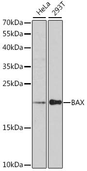
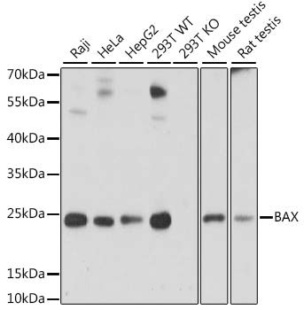
Western blot analysis of extracts of various cell lines, using BAX antibody at 1:1000 dilution.
Secondary antibody: HRP Goat Anti-Rabbit IgG (H+L) at 1:10000 dilution.
Lysates/proteins: 25ug per lane.
Blocking buffer: 3% nonfat dry milk in TBST.
Detection: ECL Enhanced Kit
Exposure time: 90s.

Western blot analysis of extracts of various cell lines, using BAX antibody at 1:1000 dilution. Secondary antibody: HRP Goat Anti-Rabbit IgG (H+L) at 1:10000 dilution. Lysates/proteins: 25ug per lane. Blocking buffer: 3% nonfat dry milk in TBST. Detection: ECL Enhanced Kit Exposure time: 90s.

Catalog Number:AB-82329
Size:100 ug
Concentration:1mg/ml
Host:Rabbit
Isotype:IgG
Clone:POLY
Immunogen:A synthetic peptide corresponding to a sequence within amino acids 1-100 of human BAX
Reactivity:Human, Mouse, Rat
Applications:Western Blot: 1:500-1:1000 Immunohistochemistry: 1:100 - 1:200 Immunofluorescence: 1:50 - 1:200 Immunoprecipitation: 1:50 - 1:100
Molecular Weight:21 kDa
Purification:Affinity Purified
Form:Liquid
Synonyms:BCL2L4

Applications

Western Blot: 1:500-1:1000 Immunohistochemistry: 1:100 - 1:200 Immunofluorescence: 1:50 - 1:200 Immunoprecipitation: 1:50 - 1:100

Immunogen

A synthetic peptide corresponding to a sequence within amino acids 1-100 of human BAX
Target Background

The protein encoded by this gene belongs to the BCL2 protein family. BCL2 family members form hetero- or homodimers and act as anti- or pro-apoptotic regulators that are involved in a wide variety of cellular activities. This protein forms a heterodimer with BCL2, and functions as an apoptotic activator. This protein is reported to interact with, and increase the opening of, the mitochondrial voltage-dependent anion channel (VDAC), which leads to the loss in membrane potential and the release of cytochrome c. The expression of this gene is regulated by the tumor suppressor P53 and has been shown to be involved in P53-mediated apoptosis. Multiple alternatively spliced transcript variants, which encode different isoforms, have been reported for this gene.

Synonyms:BCL2L4

Storage

Store at -20℃. Avoid freeze / thaw cycles.

Buffer

PBS with 0.02% sodium azide, 50% glycerol, pH7.3.

Western blot analysis of extracts of various cell lines, using BAX antibody at 1:1000 dilution. Secondary antibody: HRP Goat Anti-Rabbit IgG (H+L) at 1:10000 dilution. Lysates/proteins: 25ug per lane. Blocking buffer: 3% nonfat dry milk in TBST. Detection: ECL Enhanced Kit Exposure time: 90s.

Western blot analysis of extracts of various cell lines, using BAX antibody at 1:1000 dilution. Secondary antibody: HRP Goat Anti-Rabbit IgG (H+L) at 1:10000 dilution. Lysates/proteins: 25ug per lane. Blocking buffer: 3% nonfat dry milk in TBST. Detection: ECL Basic Kit. Exposure time: 5s.

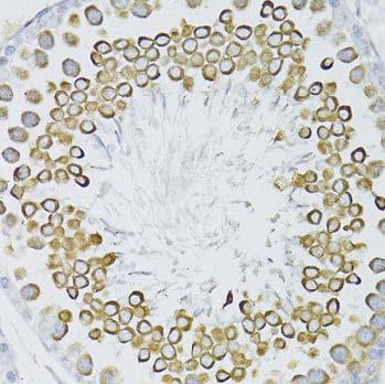
Immunohistochemistry of paraffin embedded rat testis using BAX antibody at dilution of 1:100 (40x lens).

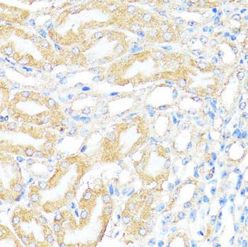
Immunohistochemistry of paraffinembedded mouse kidney using BAX antibody at dilution of 1:100 (40x lens).

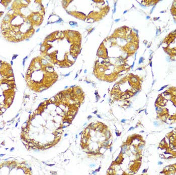
Immunohistochemistry of paraffinembedded human stomach using BAX antibody at dilution of 1:100 (40x lens).

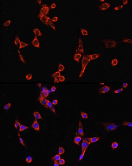
Immunofluorescence analysis of NIH/3T3 cells using BAX antibody at dilution of 1:100. Blue: DAPI for nuclear staining

No documents available.