BCL-2 Mouse Monoclonal Antibody

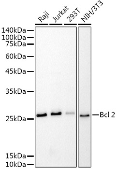
Western blot analysis of extracts of various cell lines, using Bcl-2 antibody ) at 1:1000
dilution.
Secondary antibody: HRP Goat Anti-Mouse IgG (H+L) at 1:10000 dilution.
Lysates/proteins: 25ug per lane.
Blocking buffer: 3% nonfat dry milk in TBST.
Detection: ECL West Pico plus.
Exposure time: 10s.

Western blot analysis of extracts of various cell lines, using Bcl-2 antibody ) at 1:1000 dilution. Secondary antibody: HRP Goat Anti-Mouse IgG (H+L) at 1:10000 dilution. Lysates/proteins: 25ug per lane. Blocking buffer: 3% nonfat dry milk in TBST. Detection: ECL West Pico plus. Exposure time: 10s.

Catalog Number:MAB-94760
Conjugate:Unconjugated
Size:100 ug
Concentration:1mg/ml
Host:Mouse
Isotype:IgG1
Clone:BCL2/100
Immunogen:Synthetic peptide corresponding to the amino acids 41-54 of Hu BCL2
Reactivity:Human
Applications:Western Blot:1-2 ug/ml - Immunohistochemistry: 1-10 ug/ml - FC: 1-5 ug/ml
Molecular Weight:26KDa
Purification:Affinity purification
Form:Liquid
Synonyms:Bcl-2;PPP1R50;BCL2

Applications

Western Blot:1-2 ug/ml - Immunohistochemistry: 1-10 ug/ml - FC: 1-5 ug/ml

Immunogen

Synthetic peptide corresponding to the amino acids 41-54 of Hu BCL2
Target Background

  Bcl2 (B cell chronic lymphatic leukemia protein 2) is a proto-oncogen, which can contribute to tumorigenesis by counteracting apoptosis in various cell types. The anti-apoptotic effect of Bcl2 is performed by its interactions with suppressors and agonists of cell death and under physiological conditions it is regulated by proteolytic processing and phosphorylation. Bcl2 expression can be detected mainly in lymphoid tissues and in the basal cells of epithelial tissues. It is also a marker that can help in classification of lymphoproliferative diseases and in prognostics of some epithelial neoplasms.

Synonyms:Bcl-2;PPP1R50;BCL2

Storage

Store at 2-8° for short term. Do not freeze . Avoid freeze / thaw cycles.

Buffer

Phosphate buffered saline (PBS), pH 7.4, 15 mM sodium azide

Western blot analysis of extracts of various cell lines, using Bcl-2 antibody ) at 1:1000 dilution. Secondary antibody: HRP Goat Anti-Mouse IgG (H+L) at 1:10000 dilution. Lysates/proteins: 25ug per lane. Blocking buffer: 3% nonfat dry milk in TBST. Detection: ECL West Pico plus. Exposure time: 10s.

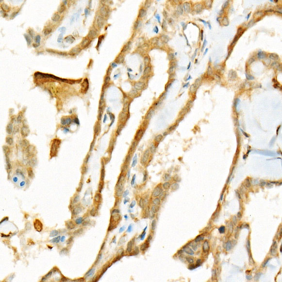
Immunohistochemistry of paraffinembedded human thyroid cancer using Bcl-2 Mouse mAb  at dilution of 1:500 (40x lens).Perform high pressure antigen retrieval with 10 mM citrate buffer pH 6.0 before commencing with IHC staining protocol.

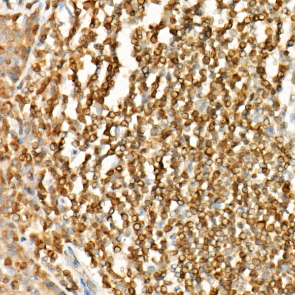
Immunohistochemistry of paraffinembedded human tonsil using Bcl-2 Mouse mAb at dilution of 1:500 (40x lens).Perform high pressure antigen retrieval with 10 mM citrate buffer pH 6.0 before commencing with IHC staining protocol.

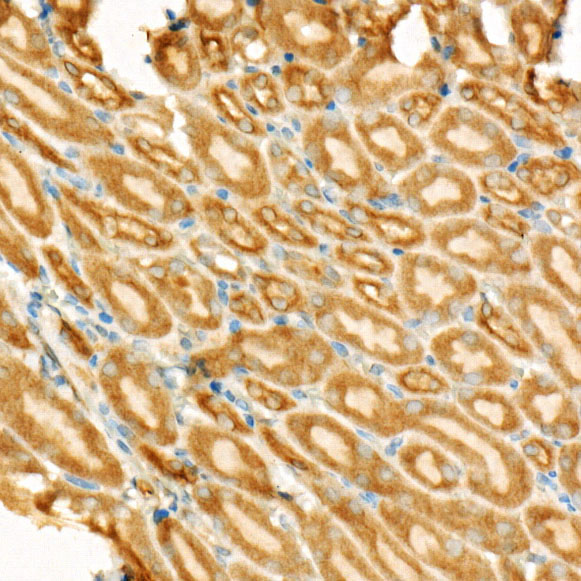
Immunohistochemistry of paraffinembedded mouse kidney using Bcl-2 Mouse mAb  at dilution of 1:500 (40x lens).Perform high pressure antigen retrieval with 10 mM citrate buffer pH 6.0 before commencing with IHC staining protocol.

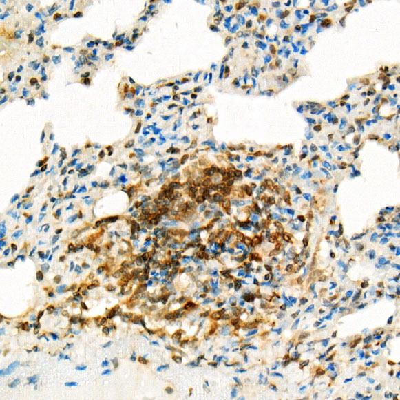
Immunohistochemistry of paraffinembedded rat lung using Bcl-2 Mouse mAb  at dilution of 1:500 (40x lens).Perform high pressure antigen retrieval with 10 mM citrate buffer pH 6.0 before commencing with IHC staining protocol.

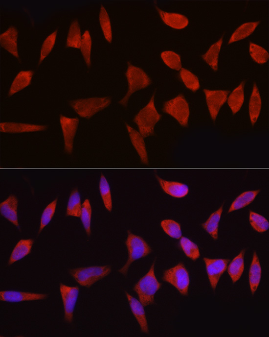
Immunofluorescence analysis of HeLa cells using Bcl-2 Mouse mAb at dilution of 1:100 (40x lens). Blue: DAPI for nuclear staining.

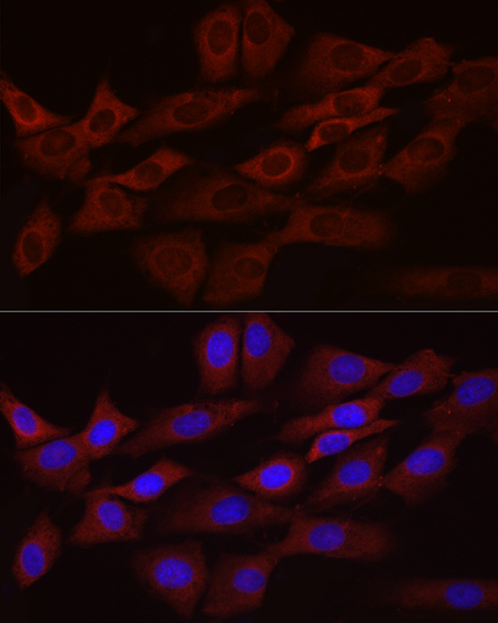
Immunofluorescence analysis of NIH/3T3 cells using Bcl-2 Mouse mAb  at dilution of 1:100 (40x lens). Blue: DAPI for nuclear staining.

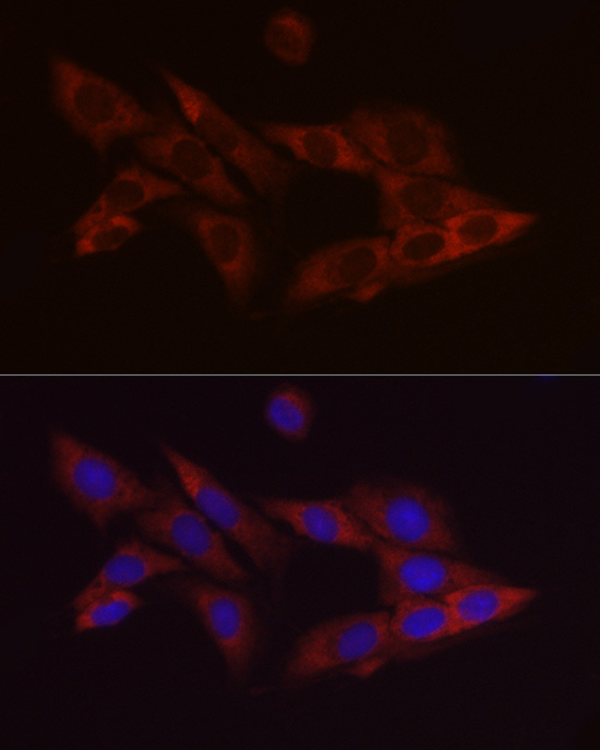
Immunofluorescence analysis of PC-12 cells using Bcl-2 Mouse mAb at dilution of 1:100 (40x lens). Blue: DAPI for nuclear staining.

No documents available.