BDNF
![Prof.Rosa Di Liddo , Dr. Thomas Bertalot, Dr. Sandra Schrenk
University of Padova
Department of Pharmaceutical and Pharmacological Sciences
See western blot protocol on Western Blotting AB-82598
(Protocol of Prof.Rosa Di Liddo,Dr.Thomas Bertalot, Dr. Sandra Schrenk)
University of Padova
Department of Pharmaceutical and Pharmacological Sciences
Sample preparation
Whole protein lysate was obtained using RIPA lysis buffer supplemented with protease inhibitors. All procedures were performed on ice and homogenization of samples was executed using a 22G needle syringe.
1. SDS-PAGE Electrophoresis: Sample loaded: 10μg - Poly-Acrylamide gel at 12%
2. Transfer: 0.45μm PVDF Membrane (Transfer for 80 min at 4°C on ice) -140 Volts and 14 mA
3. Blocking: - Blocking solution: 5% no-fat milk with TBS -Time: 2h
4. Incubation of Primary antibody: Dilution of primary antibody: 1:1000 [1μg/mL] -Dilute buffer: 1% no-fat milk with
TBS - Time and temperature: 4°C overnight in constant slow shacking
5. Washing: Wash with TBS-T (Tween-20, 0.25%) for three times (15 min, 5 min, 5 min)
6. Incubation of Secondary antibody: Dilution of secondary antibody: HRP-Goat anti-rabbit IgG (1:2500)
7. Dilute buffer: 1% no-fat milk with TBS -Time and temperature: RT, 90min on shaker
8. Washing: Wash with TBS-T (Tween-20, 0.25%) for four times (20 min, 5 min, 5 min, 5 min)
9. ECL chemiluminescent method:](/storage/products/wp-images/2022/01/AB-82598-1.jpg)
Prof.Rosa Di Liddo , Dr. Thomas Bertalot, Dr. Sandra Schrenk University of Padova Department of Pharmaceutical and Pharmacological Sciences See western blot protocol on Western Blotting AB-82598 (Protocol of Prof.Rosa Di Liddo,Dr.Thomas Bertalot, Dr. Sandra Schrenk) University of Padova Department of Pharmaceutical and Pharmacological Sciences Sample preparation Whole protein lysate was obtained using RIPA lysis buffer supplemented with protease inhibitors. All procedures were performed on ice and homogenization of samples was executed using a 22G needle syringe. 1. SDS-PAGE Electrophoresis: Sample loaded: 10μg - Poly-Acrylamide gel at 12% 2. Transfer: 0.45μm PVDF Membrane (Transfer for 80 min at 4°C on ice) -140 Volts and 14 mA 3. Blocking: - Blocking solution: 5% no-fat milk with TBS -Time: 2h 4. Incubation of Primary antibody: Dilution of primary antibody: 1:1000 [1μg/mL] -Dilute buffer: 1% no-fat milk with TBS - Time and temperature: 4°C overnight in constant slow shacking 5. Washing: Wash with TBS-T (Tween-20, 0.25%) for three times (15 min, 5 min, 5 min) 6. Incubation of Secondary antibody: Dilution of secondary antibody: HRP-Goat anti-rabbit IgG (1:2500) 7. Dilute buffer: 1% no-fat milk with TBS -Time and temperature: RT, 90min on shaker 8. Washing: Wash with TBS-T (Tween-20, 0.25%) for four times (20 min, 5 min, 5 min, 5 min) 9. ECL chemiluminescent method:
| Catalog Number: | AB-82598 |
| Conjugate: | Unconjugated |
| Size: | 100 ug |
| Concentration: | 1mg/ml |
| Host: | Rb |
| Isotype: | IgG |
| Clone: | POLY |
| Immunogen: | E.coli-derived human BDNF recombinant protein (Position: H129-R247). Human BDNF shares 100% amino acid (aa) sequence identity with both mouse and rat BDNF. |
| Reactivity: | Hu,Ms,Rt |
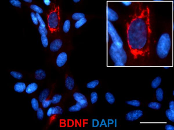
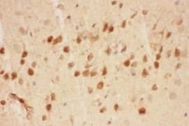
| Applications: | • Western Blot : 1:500 – 1:1000 • IHC(paraffin embedded): 1:500 – 1:1000 • IF (Immunofluorescence):1:50 |
| Molecular Weight: | 37 kDa , 25 kDa |
| Purification: | Aff. Pur. |
| Form: | Liquid |
Applications
• Western Blot : 1:500 – 1:1000 • IHC(paraffin embedded): 1:500 – 1:1000 • IF (Immunofluorescence):1:50
Immunogen
E.coli-derived human BDNF recombinant protein (Position: H129-R247). Human BDNF shares 100% amino acid (aa) sequence identity with both mouse and rat BDNF.