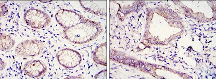
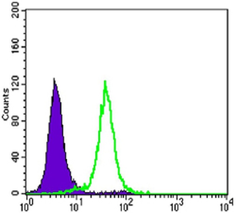
Cadherin-E
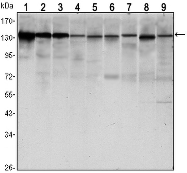
Western blot analysis using E-cadherin monoclonal antibody against LNCAP (1) , A431 (2) , DU145 (3) , PC-3 (4) , MCF-7 (5),PC-12 (6) , NIH/3T3 (7) , C6 (8) and COS7 (9) cell lysate.
| Catalog Number: | MAB-94623 |
| Conjugate: | Unconjugated |
| Size: | 100 ug |
| Concentration: | 1mg/ml |
| Host: | MS |
| Isotype: | IgG1 |
| Clone: | 7H12 |
| Immunogen: | Purified recombinant fragment of human E-cadherin expressed in E. Coli. |
| Reactivity: | Hu, Ms, Mk |
| Applications: | Western Blot: 1:500-1:2000 Immunohistochemistry: 1:200-1:1000 Immunofluorescence: 1:200-1:500 Flow Cytometry: 1:200-1:400 ELISA: 1:10000 |
| Molecular Weight: | 135kDa |
| Purification: | Aff. Pur. |
| Form: | Liquid |
Applications
Western Blot: 1:500-1:2000 Immunohistochemistry: 1:200-1:1000 Immunofluorescence: 1:200-1:500 Flow Cytometry: 1:200-1:400 ELISA: 1:10000
Immunogen
Purified recombinant fragment of human E-cadherin expressed in E. Coli.