Cadherin-E

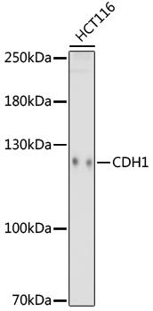
Western blot analysis of extracts of HCT116 cells, using CDH1 antibody at 1:1000 dilution.
Secondary antibody: HRP Goat Anti-Rabbit IgG (H+L) at 1:10000 dilution.
Lysates/proteins: 25ug per lane.
Blocking buffer: 3% nonfat dry milk in TBST.
Detection: ECL West Pico Plus.
Exposure time: 180s.

Western blot analysis of extracts of HCT116 cells, using CDH1 antibody at 1:1000 dilution. Secondary antibody: HRP Goat Anti-Rabbit IgG (H+L) at 1:10000 dilution. Lysates/proteins: 25ug per lane. Blocking buffer: 3% nonfat dry milk in TBST. Detection: ECL West Pico Plus. Exposure time: 180s.

Catalog Number:MAB-94628
Conjugate:Unconjugated
Size:100 ug
Concentration:1mg/ml
Host:Ms
Isotype:IgG
Clone:CN-2
Immunogen:Recombinant protein of human E-Cadherin.
Reactivity:Hu, Ms, Rt
Applications:Western Blot: 1:500 - 1:2000 Immunofluorescence: 1:50 - 1:200
Molecular Weight:120kDa
Purification:Aff. Pur.
Form:Liquid
Synonyms:Arc-1;CD324;CDHE;ECAD;LCAM;UVO;CDH1;E-Cadherin

Applications

Western Blot: 1:500 - 1:2000 Immunofluorescence: 1:50 - 1:200

Immunogen

Recombinant protein of human E-Cadherin.
Target Background

This gene encodes a classical cadherin of the cadherin superfamily. Alternative splicing results in multiple transcript variants, at least one of which encodes a preproprotein that is proteolytically processed to generate the mature glycoprotein. This calcium dependent cell-cell adhesion protein is comprised of five extracellular cadherin repeats, a transmembrane region and a highly conserved cytoplasmic tail. Mutations in this gene are correlated with gastric, breast, colorectal, thyroid and ovarian cancer. Loss of function of this gene is thought to contribute to cancer progression by increasing proliferation, invasion, and/or metastasis. The ectodomain of this protein mediates bacterial adhesion to mammalian cells and the cytoplasmic domain is required for internalization. This gene is present in a gene cluster with other members of the cadherin family on chromosome 16.

Synonyms:Arc-1;CD324;CDHE;ECAD;LCAM;UVO;CDH1;E-Cadherin

Storage

Store at -20℃. Avoid freeze / thaw cycles.

Buffer

PBS with 0.02% sodium azide,50% glycerol,pH7.3.

Western blot analysis of extracts of HCT116 cells, using CDH1 antibody at 1:1000 dilution. Secondary antibody: HRP Goat Anti-Rabbit IgG (H+L) at 1:10000 dilution. Lysates/proteins: 25ug per lane. Blocking buffer: 3% nonfat dry milk in TBST. Detection: ECL West Pico Plus. Exposure time: 180s.

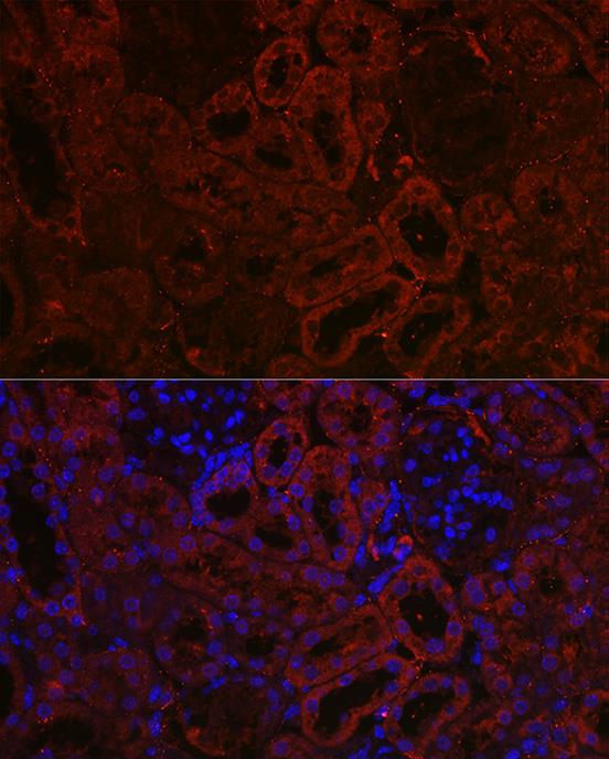
Immunofluorescence analysis of rat kidney using E-Cadherin Mouse mAb at dilution of 1:100 (40x lens). Blue: DAPI for nuclear staining.

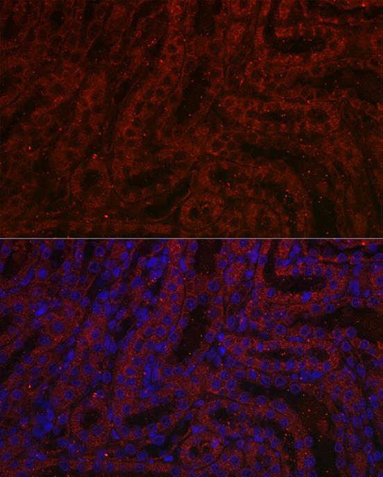
Immunofluorescence analysis of mouse kidney using E-Cadherin Mouse mAb at dilution of 1:100 (40x lens). Blue: DAPI for nuclear staining.

No documents available.