Caspase 3 (active) Mouse Monoclonal Antibody
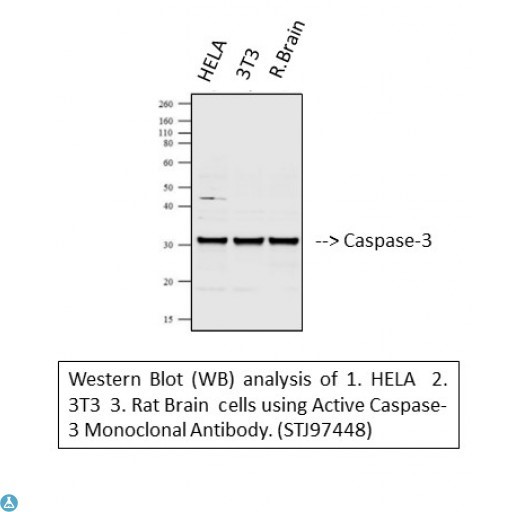
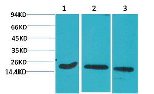
Western Blot analysis of 1. HELA 2.0  3T3 Rat Brain cells using Active Casapse-3 Monoclonal Antibody

Western Blot analysis of 1. HELA 2.0 3T3 Rat Brain cells using Active Casapse-3 Monoclonal Antibody

Catalog Number:MAB-94427
Conjugate:Unconjugated
Size:100 ug
Concentration:1mg/ml
Host:Mouse
Isotype:IgG1
Clone:5E1
Immunogen:Recombinant Protein
Reactivity:Avian, Human, Mouse, Rat
Applications:Western Blot: 1:500-1000 Immunohistochemistry: 1:100-200 Immunofluorescence: 1:100
Molecular Weight:31 kDa
Purification:The antibody was affinity-purified from mouse ascites by affinity chromatography using epitope-specific immunogen.
Form:Liquid
Synonyms:Caspase-3 CASP-3 Apopain Cysteine protease CPP32 CPP-32 Protein Yama SREBP cleavage activity 1 SCA-1 Caspase-3 subunit p17 Caspase-3 subunit p12

Applications

Western Blot: 1:500-1000 Immunohistochemistry: 1:100-200 Immunofluorescence: 1:100

Immunogen

Recombinant Protein
Target Background

Active caspase-3 is a protein encoded by the CASP3 gene which is approximately 31,6 kDa. Active caspase-3 is localised to the cytoplasm. It is involved in the TNFR1 pathway, apoptosis modulation and signaling and respiratory electron transport. This protein falls under the cysteineaspartic acid protease family. It is a sequential activator of caspases and plays a central role in the execution-phase of cell apoptosis. Caspases exist as inactive proenzymes which undergo proteolytic processing at conserved aspartic residues to produce two subunits, large and small, that dimerize to form the active enzyme. Active caspase-3 is highly expressed in lung, spleen, heart, liver and kidney. Mutations in the CASP3 gene may result in bladder urothelial carcinoma and primary effusion lymphoma. Active Caspase 3 was affinity-purified from mouse ascites by affinity-chromatography using epitope-specific immunogen. This antibody detects endogenous active caspase-3 protein.

Synonyms:Caspase-3 CASP-3 Apopain Cysteine protease CPP32 CPP-32 Protein Yama SREBP cleavage activity 1 SCA-1 Caspase-3 subunit p17 Caspase-3 subunit p12

Storage

Store at -20°C, and avoid repeat freeze-thaw cycles.

Buffer

Liquid in PBS containing 50% glycerol, 0.5% BSA and 0.02% sodium azide.

Western Blot analysis of 1. HELA 2.0 3T3 Rat Brain cells using Active Casapse-3 Monoclonal Antibody

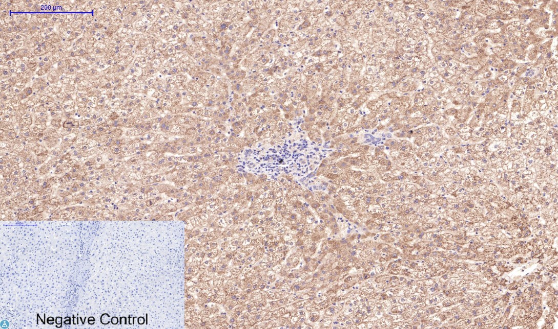
Immunohistochemical analysis of human-liver tissue. Anti-Active Caspase-3 at 1:200 (4°C, overnight). Antigen retrieval - Sodium Citrate pH6 (>98°C, 20min). Secondary - 1:200 (room temp, 30min). Negative control - Secondary only

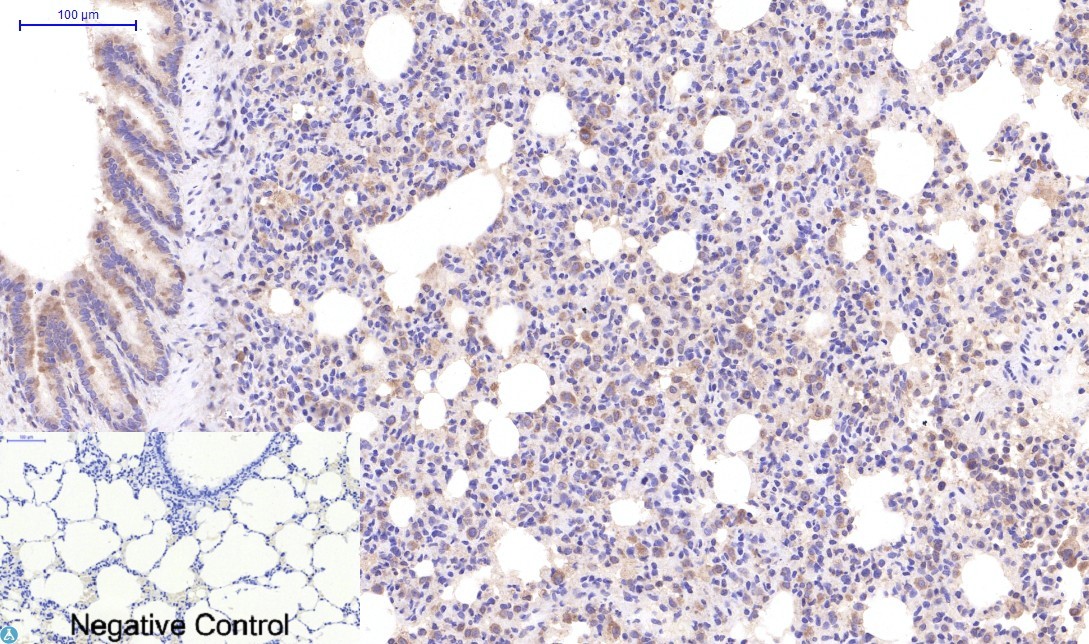
Immunohistochemical analysis of mouse lung tissue. Anti-Active Caspase-3 at 1:200 (4°C, overnight). Antigen retrieval - Sodium Citrate pH6 (>98°C, 20min). Secondary - 1:200 (room temp, 30min). Negative control - Secondary only

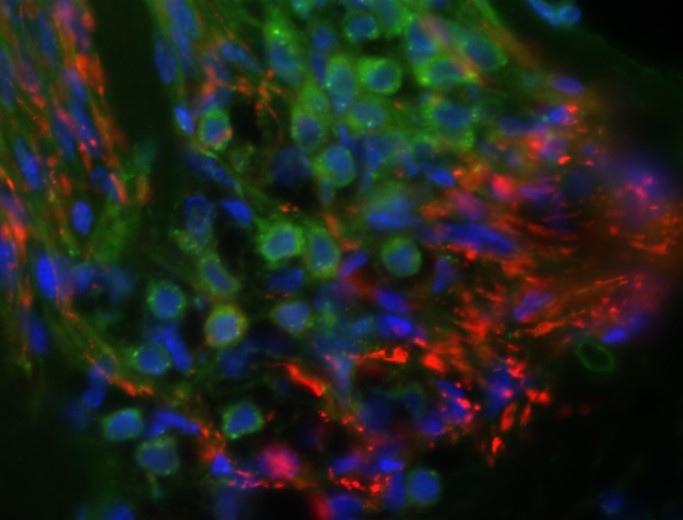
Application Immunofluorescence Antibody Name Anti-cleaved-caspase-3 Primary Dilution 1:100 Primary Probing for 2’ at 13krpm to remove clumps and applied to sections. Slides are left o/n at 4C Secondary Antibody Anti-rabbit alexa 568 Reference Antibody N/A Secondary Dilution 1:400 Secondary Probing RT for 2h – alexa 568 and alexa489 Tissue Cochlea Tissue Processing 30% and then embedded in OCT. Sections are cut at 12um onto gelatin-subbed slides and stored at -20C. Positive Control N/A Negative Control N/A Blocking 1-2h in 5% donkey serum/1% BSA in PBS/triton. Wash several times for 5’ each in PBS triton Visualization Sections are mounted in a fluorescence-protective medium Specificity 3 Findings 1:100 seems to be okay, doesn’t give too much background staining

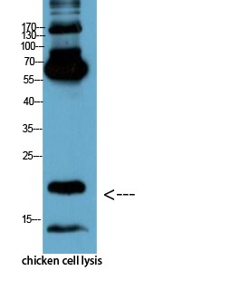
Western Blot analysis of chicken cell lysis using Antibody diluted at 1:1000

Western blot analysis of 1) Hela, 2) 3T3, 3) Rat Brain Tissue using Active Caspase-3 Monoclonal Antibody

No documents available.