Caspase 9

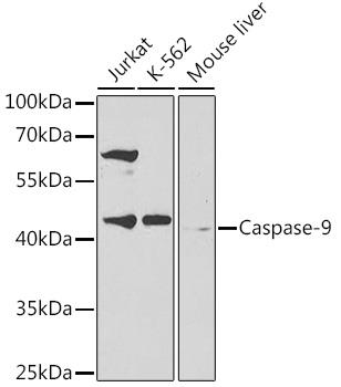
Western blot analysis of extracts of various cell lines, using Caspase-9 antibody  at 1:300
dilution.
Secondary antibody: HRP Goat Anti-Rabbit IgG (H+L) at 1:10000 dilution.
Lysates/proteins: 25ug per lane.
Blocking buffer: 3% nonfat dry milk in TBST.
Detection: ECL West Pico Plus.   
Exposure time: 60s.

Western blot analysis of extracts of various cell lines, using Caspase-9 antibody at 1:300 dilution. Secondary antibody: HRP Goat Anti-Rabbit IgG (H+L) at 1:10000 dilution. Lysates/proteins: 25ug per lane. Blocking buffer: 3% nonfat dry milk in TBST. Detection: ECL West Pico Plus. Exposure time: 60s.

Catalog Number:AB-82859
Conjugate:Unconjugated
Size:100 ug
Concentration:1mg/ml
Host:Rabbit
Isotype:IgG
Clone:POLY
Immunogen:Recombinant protein of human Caspase-9
Reactivity:Ms, Rt, Hu
Applications:Western Blot: 1:500 - 1:2000 Immunohistochemistry: 1:50 - 1:200 Immunofluorescence: 1:50 - 1:100
Molecular Weight:36kDa/46kDa
Purification:Affinity purification
Form:Liquid
Synonyms:CASP9;APAF-3;APAF3;ICE-LAP6;MCH6;PPP1R56;caspase-9;casp9;Caspase 9

Applications

Western Blot: 1:500 - 1:2000 Immunohistochemistry: 1:50 - 1:200 Immunofluorescence: 1:50 - 1:100

Immunogen

Recombinant protein of human Caspase-9
Target Background

This gene encodes a member of the cysteine-aspartic acid protease (caspase) family. Sequential activation of caspases plays a central role in the execution-phase of cell apoptosis. Caspases exist as inactive proenzymes which undergo proteolytic processing at conserved aspartic residues to produce two subunits, large and small, that dimerize to form the active enzyme. This protein can undergo autoproteolytic processing and activation by the apoptosome, a protein complex of cytochrome c and the apoptotic peptidase activating factor 1; this step is thought to be one of the earliest in the caspase activation cascade. This protein is thought to play a central role in apoptosis and to be a tumor suppressor. Alternative splicing results in multiple transcript variants.

Synonyms:CASP9;APAF-3;APAF3;ICE-LAP6;MCH6;PPP1R56;caspase-9;casp9;Caspase 9

Storage

Store at -20°C, and avoid repeat freeze-thaw cycles.

Buffer

PBS with 0.02% sodium azide and 50% glycerol pH 7.4

Western blot analysis of extracts of various cell lines, using Caspase-9 antibody at 1:300 dilution. Secondary antibody: HRP Goat Anti-Rabbit IgG (H+L) at 1:10000 dilution. Lysates/proteins: 25ug per lane. Blocking buffer: 3% nonfat dry milk in TBST. Detection: ECL West Pico Plus. Exposure time: 60s.

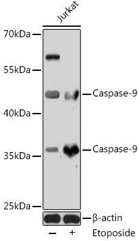
Western blot analysis of extracts of Jurkat cells, using Caspase-9 antibody at 1:1000 dilution.Jurkat cells were treated by Etoposide (25 uM) at 37℃ for 5 hours. Secondary antibody: HRP Goat Anti-Rabbit IgG (H+L) (AS014) at 1:10000 dilution. Lysates/proteins: 25ug per lane. Blocking buffer: 3% nonfat dry milk in TBST. Detection: ECL West Pico Plus. Exposure time: 30s.

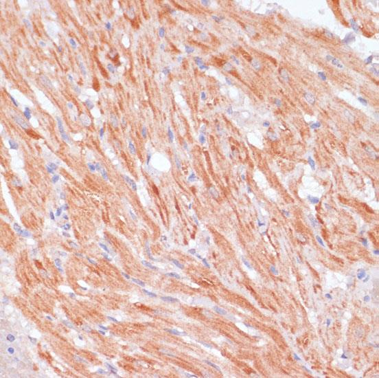
Immunohistochemistry of paraffin embedded rat heart using Caspase-9 antibody at dilution of 1:100 (40x lens).

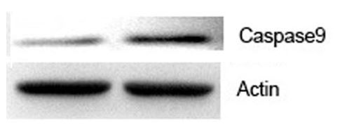
Caspase-9 Rabbit pAb5 Experiment Type Western blot(WB)

No documents available.