
Catalase

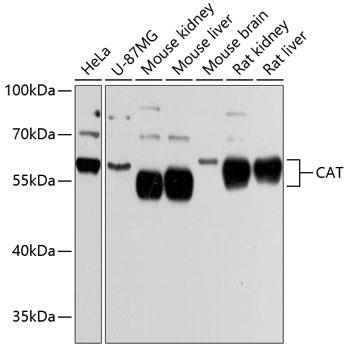
Western blot analysis of extracts of various cell lines, using CAT Antibody at 1:3000 dilution.
Secondary antibody: HRP Goat Anti-Rabbit IgG (H+L) at 1:10000 dilution.
Lysates/proteins: 25ug per lane.
Blocking buffer: 3% nonfat dry milk in TBST.
Detection: ECL West Pico Plus.
Exposure time: 5s.

Western blot analysis of extracts of various cell lines, using CAT Antibody at 1:3000 dilution. Secondary antibody: HRP Goat Anti-Rabbit IgG (H+L) at 1:10000 dilution. Lysates/proteins: 25ug per lane. Blocking buffer: 3% nonfat dry milk in TBST. Detection: ECL West Pico Plus. Exposure time: 5s.

Catalog Number:MAB-94599
Size:100ug
Concentration:1mg/ml
Host:Rb
Isotype:IgG
Clone:C-28
Immunogen:Recombinant fusion protein containing a sequence corresponding to amino acids 1-225 of human CAT.
Reactivity:Hu, Ms, Rt
Applications:Western Blot: 1:500 - 1:2000 Immunohistochemistry: 1:50 - 1:100 Immunofluorescence: 1:50 - 1:100
Molecular Weight:60kDa
Purification:Aff. Pur.
Form:Liquid
Synonyms:CAT;catalase;MGC138422;MGC138424

Applications

Western Blot: 1:500 - 1:2000 Immunohistochemistry: 1:50 - 1:100 Immunofluorescence: 1:50 - 1:100

Immunogen

Recombinant fusion protein containing a sequence corresponding to amino acids 1-225 of human CAT.
Target Background

This gene encodes catalase, a key antioxidant enzyme in the bodies defense against oxidative stress. Catalase is a heme enzyme that is present in the peroxisome of nearly all aerobic cells. Catalase converts the reactive oxygen species hydrogen peroxide to water and oxygen and thereby mitigates the toxic effects of hydrogen peroxide. Oxidative stress is hypothesized to play a role in the development of many chronic or late-onset diseases such as diabetes, asthma, Alzheimer's disease, systemic lupus erythematosus, rheumatoid arthritis, and cancers. Polymorphisms in this gene have been associated with decreases in catalase activity but, to date, acatalasemia is the only disease known to be caused by this gene.

Synonyms:CAT;catalase;MGC138422;MGC138424

Storage

Storage-20℃ for 12 months (Avoid repeated freeze / thaw cycles.) T483

Buffer

PBS with 0.02% sodium azide and 50% glycerol pH 7.3

Western blot analysis of extracts of various cell lines, using CAT Antibody at 1:3000 dilution. Secondary antibody: HRP Goat Anti-Rabbit IgG (H+L) at 1:10000 dilution. Lysates/proteins: 25ug per lane. Blocking buffer: 3% nonfat dry milk in TBST. Detection: ECL West Pico Plus. Exposure time: 5s.

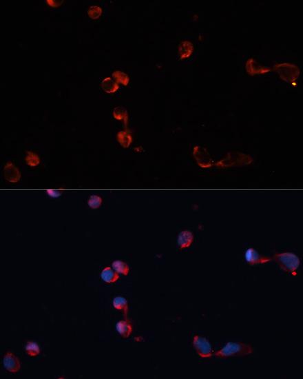
Immunofluorescence analysis of HeLa cells using CAT antibody at dilution of 1:100. Blue: DAPI for nuclear staining.

No documents available.