Catenin beta-1/CTNNB1

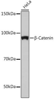
Western blot analysis of extracts of HeLa cells, using β-Catenin antibody at 1:1000 dilution. Secondary antibody: HRP Goat Anti-Rabbit IgG (H+L) at 1:10000 dilution.
Lysates/proteins: 25ug per lane. Blocking buffer: 3% nonfat dry milk in TBST.
Detection: ECL West Pico Plus. Exposure time: 10s.

Western blot analysis of extracts of HeLa cells, using β-Catenin antibody at 1:1000 dilution. Secondary antibody: HRP Goat Anti-Rabbit IgG (H+L) at 1:10000 dilution. Lysates/proteins: 25ug per lane. Blocking buffer: 3% nonfat dry milk in TBST. Detection: ECL West Pico Plus. Exposure time: 10s.

Catalog Number:MAB-94660
Size:100 ug
Concentration:1mg/ml
Host:Ms
Isotype:IgG
Clone:CB22
Immunogen:A synthesized peptide derived from human beta Catenin.
Reactivity:Hu, Ms, Rt
Applications:Western Blot: 1:500 - 1:2000 Immunohistochemistry: 1:50 - 1:200 Immunofluorescence: 1:50 - 1:200 Immunoprecipitation: 1:50 - 1:200
Molecular Weight:92kDa
Purification:Aff. Pur.
Form:Liquid
Synonyms:CTNNB;MRD19;armadillo;beta Catenin;CTNNB1

Applications

Western Blot: 1:500 - 1:2000 Immunohistochemistry: 1:50 - 1:200 Immunofluorescence: 1:50 - 1:200 Immunoprecipitation: 1:50 - 1:200

Immunogen

A synthesized peptide derived from human beta Catenin.
Target Background

The protein encoded by this gene is part of a complex of proteins that constitute adherens junctions (AJs). AJs are necessary for the creation and maintenance of epithelial cell layers by regulating cell growth and adhesion between cells. The encoded protein also anchors the actin cytoskeleton and may be responsible for transmitting the contact inhibition signal that causes cells to stop dividing once the epithelial sheet is complete. Finally, this protein binds to the product of the APC gene, which is mutated in adenomatous polyposis of the colon. Mutations in this gene are a cause of colorectal cancer (CRC), pilomatrixoma (PTR), medulloblastoma (MDB), and ovarian cancer. Alternative splicing results in multiple transcript variants.

Synonyms:CTNNB;MRD19;armadillo;beta Catenin;CTNNB1

Storage

Store at -20℃. Avoid freeze / thaw cycles.

Buffer

PBS with 0.02% sodium azide and 50% glycerol pH 7.4

Western blot analysis of extracts of HeLa cells, using β-Catenin antibody at 1:1000 dilution. Secondary antibody: HRP Goat Anti-Rabbit IgG (H+L) at 1:10000 dilution. Lysates/proteins: 25ug per lane. Blocking buffer: 3% nonfat dry milk in TBST. Detection: ECL West Pico Plus. Exposure time: 10s.

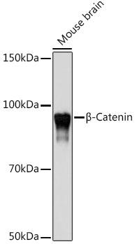
Western blot analysis of extracts of Mouse brain, using β-Catenin antibody at 1:1000 dilution. Secondary antibody: HRP Goat Anti-Rabbit IgG (H+L) at 1:10000 dilution. Lysates/proteins: 25ug per lane.Blocking buffer: 3% nonfat dry milk in TBST. Detection: ECL West Pico Plus. Exposure time: 10s.

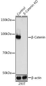
Western blot analysis of extracts from normal (control) and β-Catenin knockout (KO) 293T cells, using β-Catenin antibody at 1:1000 dilution. Secondary antibody: HRP Goat Anti-Rabbit IgG (H+L) at 1:10000 dilution. Lysates/proteins: 25ug per lane. Blocking buffer: 3% nonfat dry milk in TBST. Detection: ECL West Pico Plus. Exposure time: 10s.

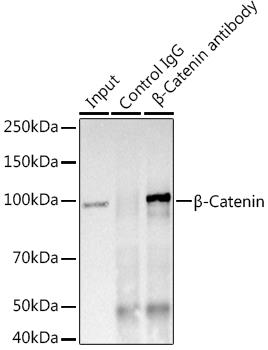
Immunoprecipitation analysis of 600ug extracts of Mouse brain using 3ug β-Catenin antibody. Western blot was performed from the immunoprecipitate using β-Catenin at a dilition of 1:1000.

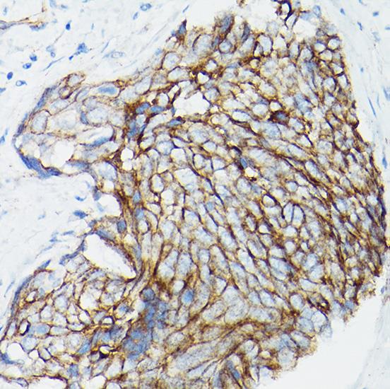
Immunohistochemistry of paraffin embedded Human cervical squamous cell carcinoma using [KO Validated] β-Catenin Rabbit mAb at dilution of 1:100 (40x lens).

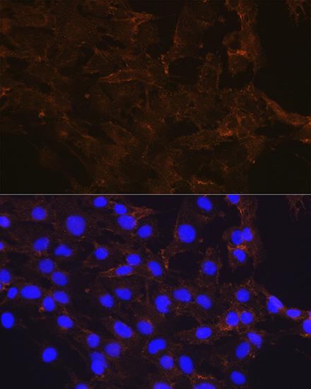
Immunofluorescence analysis of C6 cells using β-Catenin antibody at dilution of 1:100. Blue: DAPI for nuclear staining.

No documents available.