Cathepsin L Rabbit Monoclonal Antibody

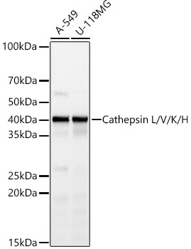
Western blot analysis of various lysates, using Cathepsin L/V/K/H Rabbit mAbat 1:1000 dilution.
Secondary antibody: HRP Goat Anti-Rabbit IgG (H+L) at 1:10000 dilution.
Lysates/proteins: 25μg per lane.
Blocking buffer: 3% nonfat dry milk in TBST.
Detection: ECL West Pico Plus.
Exposure time: 10s.

Western blot analysis of various lysates, using Cathepsin L/V/K/H Rabbit mAbat 1:1000 dilution. Secondary antibody: HRP Goat Anti-Rabbit IgG (H+L) at 1:10000 dilution. Lysates/proteins: 25μg per lane. Blocking buffer: 3% nonfat dry milk in TBST. Detection: ECL West Pico Plus. Exposure time: 10s.

Catalog Number:MAB-95047
Conjugate:Unconjugated
Size:100 ug
Concentration:1mg/ml
Host:Rabbit
Isotype:IgG
Clone:ARC59023
Immunogen:Recombinant fusion protein containing a sequence corresponding to amino acids 100-200 of human Cathepsin L
Reactivity:Human
Applications:Western Blot:   1:500 - 1:1000 Immunohistochemistry (paraffin-embedded tissues):   1:50 - 1:200
Molecular Weight:42kDa
Purification:Affinity purification
Form:Liquid
Synonyms:MEP; CATL; CTSL1; Cathepsin L

Applications

Western Blot:   1:500 - 1:1000 Immunohistochemistry (paraffin-embedded tissues):   1:50 - 1:200

Immunogen

Recombinant fusion protein containing a sequence corresponding to amino acids 100-200 of human Cathepsin L

Target Background

The protein encoded by this gene is a lysosomal cysteine proteinase that plays a major role in intracellular protein catabolism. Its substrates include collagen and elastin, as well as alpha-1 protease inhibitor, a major controlling element of neutrophil elastase activity. The encoded protein has been implicated in several pathologic processes, including myofibril necrosis in myopathies and in myocardial ischemia, and in the renal tubular response to proteinuria. This protein, which is a member of the peptidase C1 family, is a dimer composed of disulfide-linked heavy and light chains, both produced from a single protein precursor. Additionally, this protein cleaves the S1 subunit of the SARS-CoV-2 spike protein, which is necessary for entry of the virus into the cell.

Synonyms:MEP; CATL; CTSL1; Cathepsin L

Storage

Store at -20℃. Avoid freeze / thaw cycles.

Buffer

PBS with 0.05% proclin300,0.05% BSA,50% glycerol,pH7.3.

Western blot analysis of various lysates, using Cathepsin L/V/K/H Rabbit mAbat 1:1000 dilution. Secondary antibody: HRP Goat Anti-Rabbit IgG (H+L) at 1:10000 dilution. Lysates/proteins: 25μg per lane. Blocking buffer: 3% nonfat dry milk in TBST. Detection: ECL West Pico Plus. Exposure time: 10s.

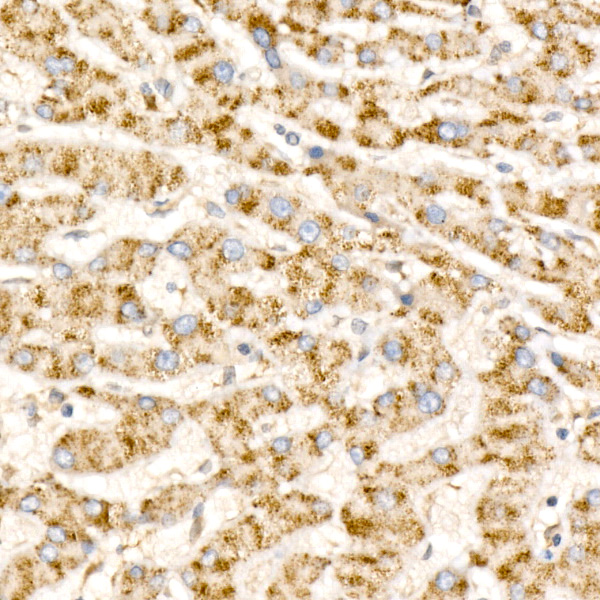
Immunohistochemistry analysis of paraffin-embedded Human liver cancer using Cathepsin L/V/K/H Rabbit mAb at dilution of 1:100 (40x lens). High pressure antigen retrieval performed with 0.01M Citrate Bufferr (pH 6.0) prior to IHC staining.

No documents available.