Cathepsin L Rabbit Polyclonal Antibody

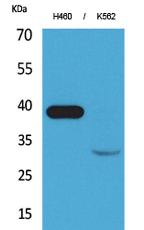
Western Blot analysis of H460, K562 cells using Cathepsin L Polyclonal Antibody. Secondary antibody was diluted at 1:20000

Western Blot analysis of H460, K562 cells using Cathepsin L Polyclonal Antibody. Secondary antibody was diluted at 1:20000

Catalog Number:AB-E4833
Conjugate:Unconjugated
Size:100 ug
Concentration:1mg/ml
Host:Rabbit
Isotype:IgG
Clone:POLY
Immunogen:Synthesized peptide derived from the Internal region of human Cathepsin L.
Reactivity:Human;Rat;Mouse;
Applications:Western Blot: 1/500 - 1/2000 Immunohistochemistry(paraffin-embedded tissues): 1:100-300
Molecular Weight:37kD
Purification:The antibody was affinity-purified from rabbit antiserum by affinity-chromatography using epitope-specific immunogen.
Form:Liquid
Synonyms:CTSL1; CTSL; Cathepsin L1; Major excreted protein; MEP

Applications

Western Blot: 1/500 - 1/2000 Immunohistochemistry(paraffin-embedded tissues): 1:100-300

Immunogen

Synthesized peptide derived from the Internal region of human Cathepsin L.
Target Background

The protein encoded by this gene is a lysosomal cysteine proteinase that plays a major role in intracellular protein catabolism. Its substrates include collagen and elastin, as well as alpha-1 protease inhibitor, a major controlling element of neutrophil elastase activity. The encoded protein has been implicated in several pathologic processes, including myofibril necrosis in myopathies and in myocardial ischemia, and in the renal tubular response to proteinuria. This protein, which is a member of the peptidase C1 family, is a dimer composed of disulfide-linked heavy and light chains, both produced from a single protein precursor. Multiple alternatively spliced transcript variants have been found for this gene.

Synonyms:CTSL1; CTSL; Cathepsin L1; Major excreted protein; MEP

Storage

Store at -20°C. Avoid repeated freeze-thaw cycles.

Buffer

Liquid in PBS containing 50% glycerol, 0.5% BSA and 0.02% sodium azide.

Western Blot analysis of H460, K562 cells using Cathepsin L Polyclonal Antibody. Secondary antibody was diluted at 1:20000

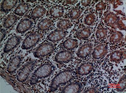
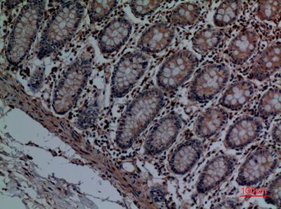
Immunohistochemical analysis of paraffin-embedded human-colon, antibody was diluted at 1:100

Immunohistochemical analysis of paraffin-embedded human-colon, antibody was diluted at 1:100

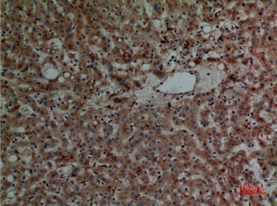
Immunohistochemical analysis of paraffin-embedded human-liver, antibody was diluted at 1:100

No documents available.