Caveolin 1 (CAV1) Rabbit Polyclonal Antibody
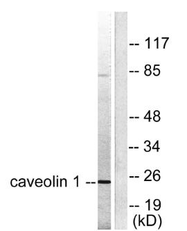
Western blot analysis of lysates from HUVEC cells, using Caveolin-1 Antibody. The lane on the right is blocked
with the synthesized peptide.

Western blot analysis of lysates from HUVEC cells, using Caveolin-1 Antibody. The lane on the right is blocked with the synthesized peptide.

Catalog Number:AB-81830
Conjugate:Unconjugated
Size:100 ug
Concentration:1mg/ml
Host:Rabbit
Isotype:IgG
Clone:POLY
Immunogen:The antiserum was produced against synthesized peptide derived from human Caveolin-1. AA range:129-178
Reactivity:Human;Mouse;Rat
Applications:Western Blot: 1:500 - 1:2000 Immunohistochemistry: 1:50 - 1:300 Immunofluorescence: 1:50 - 1:300  
Molecular Weight:25kD
Purification:The antibody was affinity-purified from rabbit antiserum by affinity-chromatography using epitope-specific immunogen.
Form:Liquid
Synonyms:CAV1; CAV; Caveolin-1

Applications

Western Blot: 1:500 - 1:2000 Immunohistochemistry: 1:50 - 1:300 Immunofluorescence: 1:50 - 1:300  

Immunogen

The antiserum was produced against synthesized peptide derived from human Caveolin-1. AA range:129-178

Target Background

The scaffolding protein encoded by this gene is the main component of the caveolae plasma membranes found in most cell types. The protein links integrin subunits to the tyrosine kinase FYN, an initiating step in coupling integrins to the Ras-ERK pathway and promoting cell cycle progression. The gene is a tumor suppressor gene candidate and a negative regulator of the Ras-p42/44 mitogen-activated kinase cascade. Caveolin 1 and caveolin 2 are located next to each other on chromosome 7 and express colocalizing proteins that form a stable hetero-oligomeric complex. Mutations in this gene have been associated with Berardinelli-Seip congenital lipodystrophy. Alternatively spliced transcripts encode alpha and beta isoforms of caveolin 1.

Synonyms:CAV1; CAV; Caveolin-1

Storage

Store at -20°C. Avoid repeated freeze-thaw cycles.

Buffer

Liquid in PBS containing 50% glycerol, 0.5% BSA and 0.02% sodium azide.

Western blot analysis of lysates from HUVEC cells, using Caveolin-1 Antibody. The lane on the right is blocked with the synthesized peptide.

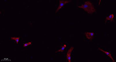
Immunofluorescence analysis of A549. 1,primary Antibody(red) was diluted at 1:200(4°C overnight). 2, Goat Anti Rabbit IgG (H&L) - Alexa Fluor 594 Secondary antibody was diluted at 1:1000(room temperature,50min).3, Picture B: DAPI(blue) 10min.

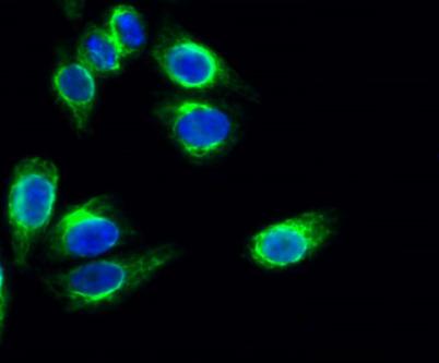
Immunofluorescence analysis of Hela cell. 1,Caveolin-1Polyclonal Antibody(green) was diluted at 1:200(4°overnight). 2, Goat Anti Rabbit Alexa Fluor 488 was diluted at 1:1000(room temperature,50min). 3 DAPI(blue) 10min.

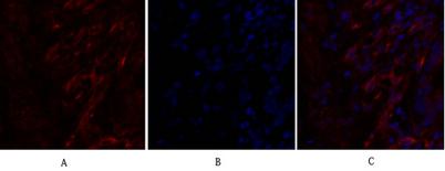
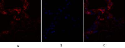
Immunofluorescence analysis of human-lung tissue.1,Caveolin-1 Polyclonal Antibody(red) was diluted at1:200(4°C,overnight). 2, Cy3 labled Secondary antibody was diluted at 1:300(room temperature, 50min).3, PictureB: DAPI(blue) 10min. Picture A:Target.

Immunofluorescence analysis of human-lung tissue.1,Caveolin-1 Polyclonal Antibody(red) was diluted at1:200(4°C,overnight). 2, Cy3 labled Secondary antibody was diluted at 1:300(room temperature, 50min).3, Picture B: DAPI(blue) 10min. Picture A:Target. Picture B: DAPI. Picture C: merge of A+B

No documents available.