
CCR2

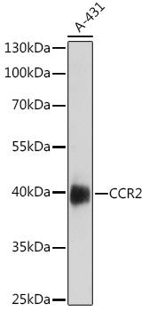
Western blot analysis of extracts of A-431 cells, using CCR2 antibody at 1:1000 dilution.
Secondary antibody: HRP Goat Anti-Rabbit IgG (H+L) at 1:10000 dilution.
Lysates/proteins: 25ug per lane.
Blocking buffer: 3% nonfat dry milk in TBST.Detection: ECL West Pico Plus.   
Exposure time: 90s.

Western blot analysis of extracts of A-431 cells, using CCR2 antibody at 1:1000 dilution. Secondary antibody: HRP Goat Anti-Rabbit IgG (H+L) at 1:10000 dilution. Lysates/proteins: 25ug per lane. Blocking buffer: 3% nonfat dry milk in TBST.Detection: ECL West Pico Plus. Exposure time: 90s.

Catalog Number:AB-84344
Conjugate:Unconjugated
Size:100ug
Concentration:1mg/ml
Host:Rb
Isotype:IgG
Clone:POLY
Immunogen:A synthetic peptide of human CCR2.
Reactivity:Hu, Ms
Applications:Western Blot: 1:500 - 1:2000 Immunofluorescence: 1:50 - 1:200
Molecular Weight:41kDa
Purification:Aff. Pur.
Form:Liquid
Synonyms:CCR2;CC-CKR-2;CCR-;CCR2A;CCR2B;CD192;CKR2;CKR2A;CKR2B;CMKBR2;MCP-1-R;FLJ78302

Applications

Western Blot: 1:500 - 1:2000 Immunofluorescence: 1:50 - 1:200

Immunogen

A synthetic peptide of human CCR2.
Target Background

This gene encodes two isoforms of a receptor for monocyte chemoattractant protein-1, a chemokine which specifically mediates monocyte chemotaxis. Monocyte chemoattractant protein-1 is involved in monocyte infiltration in inflammatory diseases such as rheumatoid arthritis as well as in the inflammatory response against tumors. The receptors encoded by this gene mediate agonist-dependent calcium mobilization and inhibition of adenylyl cyclase. This gene is located in the chemokine receptor gene cluster region. Two alternatively spliced transcript variants are expressed by the gene.

Synonyms:CCR2;CC-CKR-2;CCR-;CCR2A;CCR2B;CD192;CKR2;CKR2A;CKR2B;CMKBR2;MCP-1-R;FLJ78302

Storage

Store at -20℃. Avoid freeze / thaw cycles.

Buffer

PBS with 0.02% sodium azide,50% glycerol,pH7.3.

Western blot analysis of extracts of A-431 cells, using CCR2 antibody at 1:1000 dilution. Secondary antibody: HRP Goat Anti-Rabbit IgG (H+L) at 1:10000 dilution. Lysates/proteins: 25ug per lane. Blocking buffer: 3% nonfat dry milk in TBST.Detection: ECL West Pico Plus. Exposure time: 90s.

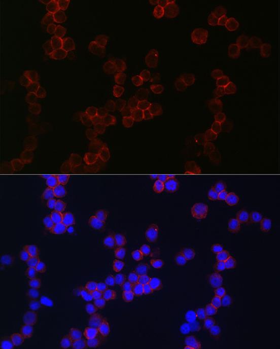
Immunofluorescence analysis of THP-1cells using CCR2 Rabbit pAb at dilution of 1:150 (40x lens). Blue: DAPI for nuclear staining.

No documents available.