CD3 epsilon Rabbit Polyclonal Antibody

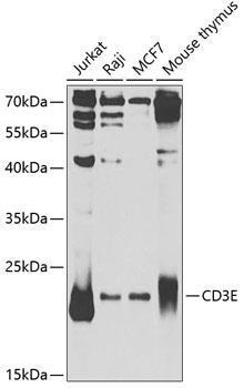
Western blot analysis of extracts of
various cell lines, using CD3E
Antibody at 1:1000 dilution.
Secondary antibody: HRP Goat Anti-
Rabbit IgG (H+L) at 1:10000
dilution.
Lysates/proteins: 25ug per lane.
Blocking buffer: 3% nonfat dry milk in
TBST.
Detection: ECL West Pico Plus 
Exposure time: 5s

Western blot analysis of extracts of various cell lines, using CD3E Antibody at 1:1000 dilution. Secondary antibody: HRP Goat Anti- Rabbit IgG (H+L) at 1:10000 dilution. Lysates/proteins: 25ug per lane. Blocking buffer: 3% nonfat dry milk in TBST. Detection: ECL West Pico Plus Exposure time: 5s

Catalog Number:AB-81831
Size:100 ug
Concentration:1mg/ml
Host:Rabbit
Isotype:IgG
Clone:POLY
Immunogen:RThe antibody was affinity-purified from rabbit antiserum by affinity-chromatography using epitope-specific immunogen.
Reactivity:Hu,Ms,Rt
Applications:Western Blot: 1:500 - 1:2000 Immunohistochemistry: 1:50 - 1:300 Immunoprecipitation: 1:50 - 1:100
Molecular Weight:21kDa
Purification:Aff. Pur.
Form:Liquid
Synonyms:CD3E;IMD18;T3E;TCRE;CD3e molecule

Applications

Western Blot: 1:500 - 1:2000 Immunohistochemistry: 1:50 - 1:300 Immunoprecipitation: 1:50 - 1:100

Immunogen

RThe antibody was affinity-purified from rabbit antiserum by affinity-chromatography using epitope-specific immunogen.

Target Background

The protein encoded by this gene is the CD3-epsilon polypeptide, which together with CD3-gamma, -delta and -zeta, and the T-cell receptor alpha/beta and gamma/delta heterodimers, forms the T-cell receptor-CD3 complex. This complex plays an important role in coupling antigen recognition to several intracellular signal-transduction pathways. The genes encoding the epsilon, gamma and delta polypeptides are located in the same cluster on chromosome 11.The epsilon polypeptide plays an essential role in T-cell development. Defects in this gene cause immunodeficiency. This gene has also been linked to a susceptibility to type I diabetes in women.

Synonyms:CD3E;IMD18;T3E;TCRE;CD3e molecule

Storage

Store at -20℃. Avoid freeze / thaw cycles.

Buffer

Liquid in PBS containing 50% glycerol, 0.5% BSA and 0.02% sodium azide.

Western blot analysis of extracts of various cell lines, using CD3E Antibody at 1:1000 dilution. Secondary antibody: HRP Goat Anti- Rabbit IgG (H+L) at 1:10000 dilution. Lysates/proteins: 25ug per lane. Blocking buffer: 3% nonfat dry milk in TBST. Detection: ECL West Pico Plus Exposure time: 5s

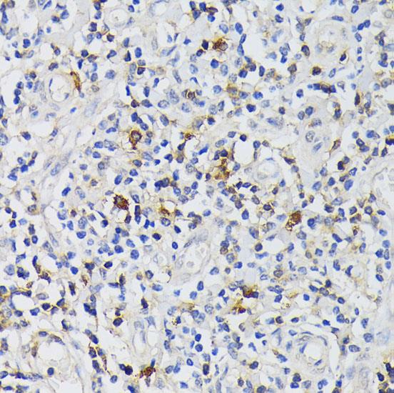
Immunohistochemistry of paraffin embedded human tonsil using CD3E Antigen antibody at dilution of 1:200 (40x lens).

No documents available.