CD31/PECAM1 Mouse Monoclonal Antibody

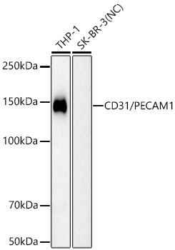
Western blot analysis of various lysates using CD31/PECAM1 Mouse  Monoclonal Ab  at 1:1000 dilution incubated overnight at 4℃.
Secondary antibody: HRP-conjugated Goat anti-Mouse IgG (H+L)  at 1:10000 dilution.
Lysates/proteins: 25 μg per lane.
Blocking buffer: 3% nonfat dry milk in TBST.
Detection: ECL Basic Kit
Negative control (NC): SK-BR-3
Exposure time: 90s.

Western blot analysis of various lysates using CD31/PECAM1 Mouse  Monoclonal Ab  at 1:1000 dilution incubated overnight at 4℃. Secondary antibody: HRP-conjugated Goat anti-Mouse IgG (H+L)  at 1:10000 dilution. Lysates/proteins: 25 μg per lane. Blocking buffer: 3% nonfat dry milk in TBST. Detection: ECL Basic Kit Negative control (NC): SK-BR-3 Exposure time: 90s.

Catalog Number:MAB-95053
Size:100 ug
Concentration:1 mg/ml
Host:Mouse
Isotype:IgG
Clone:MEM-11
Immunogen:Recombinant fusion protein containing a sequence corresponding to amino acids 630-738 of CD31/PECAM1 (
Reactivity:Human, Mouse
Applications:Western Blot 1:1000 - 1:2000 IHC-P 1:200 - 1:800 IF/ICC 1:200 - 1:800 ELISA Recommended starting concentration is 1 μg/mL.
Molecular Weight:130 kDa
Purification:Affinity Purified
Form:Liquid
Synonyms:CD31; PECA1; GPIIA'; PECAM-1; endoCAM; CD31/EndoCAM

Applications

Western Blot 1:1000 - 1:2000 IHC-P 1:200 - 1:800 IF/ICC 1:200 - 1:800 ELISA Recommended starting concentration is 1 μg/mL.

Immunogen

Recombinant fusion protein containing a sequence corresponding to amino acids 630-738 of CD31/PECAM1 (

Target Background

The protein encoded by this gene is found on the surface of platelets, monocytes, neutrophils, and some types of T-cells, and makes up a large portion of endothelial cell intercellular junctions. The encoded protein is a member of the immunoglobulin superfamily and is likely involved in leukocyte migration, angiogenesis, and integrin activation.

Synonyms:CD31; PECA1; GPIIA'; PECAM-1; endoCAM; CD31/EndoCAM

Storage

Store at -20℃. Avoid freeze / thaw cycles.

Buffer

PBS with 0.09% Sodium azide,0.05% BSA,50% glycerol,pH7.3.

Western blot analysis of various lysates using CD31/PECAM1 Mouse  Monoclonal Ab  at 1:1000 dilution incubated overnight at 4℃. Secondary antibody: HRP-conjugated Goat anti-Mouse IgG (H+L)  at 1:10000 dilution. Lysates/proteins: 25 μg per lane. Blocking buffer: 3% nonfat dry milk in TBST. Detection: ECL Basic Kit Negative control (NC): SK-BR-3 Exposure time: 90s.

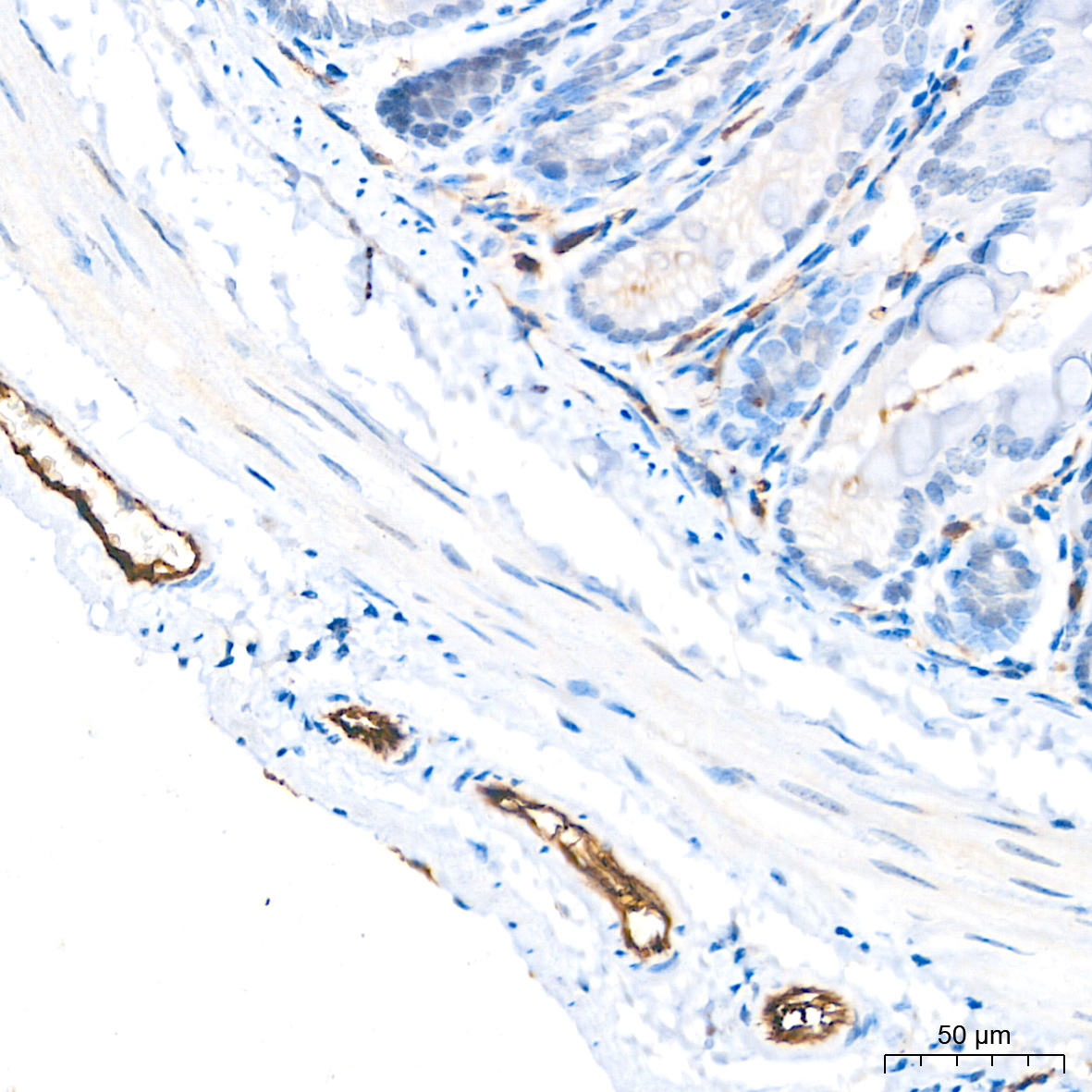
Immunohistochemistry analysis of paraffin-embedded Mouse colon tissue using CD31/PECAM1 Mouse Monoclonal Ab at a dilution of 1:200 (40x lens). High pressure antigen retrieval performed with 0.01M Citrate Buffer (pH 6.0) prior to IHC staining.

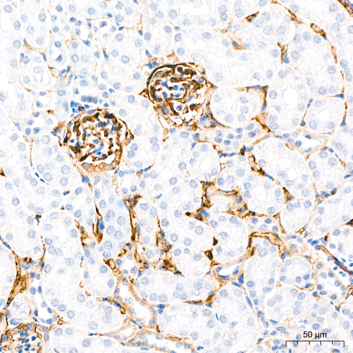
Immunohistochemistry analysis of paraffin-embedded Mouse kidney tissue using CD31/PECAM1 Mouse Monoclonal Ab at a dilution of 1:200 (40x lens). High pressure antigen retrieval performed with 0.01M Citrate Buffer (pH 6.0) prior to IHC staining.

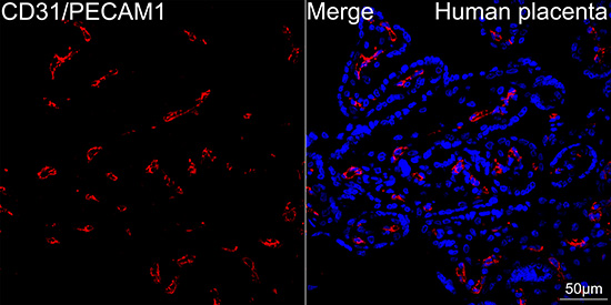
Confocal imaging of paraffinembedded Human placenta tissue using CD31/PECAM1 Mouse Monoclonal Ab at dilution 1:200 followed by a further incubation with Cy3 Goat Anti-Mouse IgG (H+L) at dilution 1:500 (Red). DAPI was used for nuclear staining (Blue). High pressure antigen retrieval performed with 0.01M Citrate Buffer (pH 6.0)

No documents available.