CD47 Rabbit Polyclonal Antibody

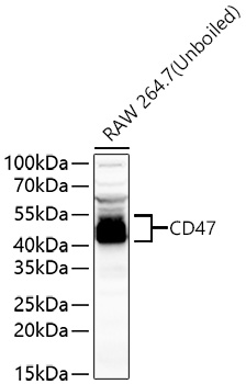
Western blot analysis of various lysates using CD47 Rabbit pAb  at 1:1000 dilution.
Secondary antibody: HRP-conjugated Goat anti-Rabbit IgG (H+L)at 1:10000 dilution.
Lysates/proteins: 25μg per lane.
Blocking buffer: 3% nonfat dry milk in TBST.
Detection: ECL West Pico Plus.   
Exposure time: 180s.

Western blot analysis of various lysates using CD47 Rabbit pAb at 1:1000 dilution. Secondary antibody: HRP-conjugated Goat anti-Rabbit IgG (H+L)at 1:10000 dilution. Lysates/proteins: 25μg per lane. Blocking buffer: 3% nonfat dry milk in TBST. Detection: ECL West Pico Plus. Exposure time: 180s.

Catalog Number:AB1838
Conjugate:Unconjugated
Size:100ug
Concentration:1mg/ml
Host:Rb
Isotype:IgG
Clone:POLY
Immunogen:A synthetic peptide corresponding to a sequence within amino acids 224-323 of human CD47 (NP_001768.1).
Reactivity:Hu,Rt,Ms,
Applications:WB,IHC,IF,ELISA
Molecular Weight:47kDa
Purification:Affinity Purified
Form:Liquid
Synonyms:None

This gene encodes a membrane protein, which is involved in the increase in intracellular calcium concentration that occurs upon cell adhesion to extracellular matrix. The encoded protein is also a receptor for the C-terminal cell binding domain of thrombospondin, and it may play a role in membrane transport and signal transduction. This gene has broad tissue distribution, and is reduced in expression on Rh erythrocytes. Alternatively spliced transcript variants have been found for this gene.

Applications

WB,IHC,IF,ELISA

Immunogen

A synthetic peptide corresponding to a sequence within amino acids 224-323 of human CD47 (NP_001768.1).

Target Background

This gene encodes a membrane protein, which is involved in the increase in intracellular calcium concentration that occurs upon cell adhesion to extracellular matrix. The encoded protein is also a receptor for the C-terminal cell binding domain of thrombospondin, and it may play a role in membrane transport and signal transduction. This gene has broad tissue distribution, and is reduced in expression on Rh erythrocytes. Alternatively spliced transcript variants have been found for this gene.

Synonyms:None

Storage

Store at -20℃. Avoid freeze / thaw cycles.

Buffer

PBS with 0.02% sodium azide,50% glycerol,pH7.3.

Western blot analysis of various lysates using CD47 Rabbit pAb at 1:1000 dilution. Secondary antibody: HRP-conjugated Goat anti-Rabbit IgG (H+L)at 1:10000 dilution. Lysates/proteins: 25μg per lane. Blocking buffer: 3% nonfat dry milk in TBST. Detection: ECL West Pico Plus. Exposure time: 180s.

Western blot analysis of lysates from U-251MG cells, using CD47 Rabbit pAb at 1:1000 dilution. Secondary antibody: HRP-conjugated Goat anti-Rabbit IgG (H+L) at 1:10000 dilution. Lysates/proteins: 25μg per lane. Blocking buffer: 3% nonfat dry milk in TBST. Detection: ECL West Pico Plus. Exposure time: 10s.

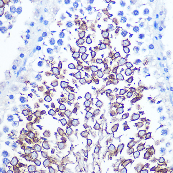
Immunohistochemistry analysis of paraffin-embedded Rat testis using CD47 Rabbit pAb at dilution of 1:100 (40x lens). Microwave antigen retrieval performed with 0.01M PBS Buffer (pH 7.2) prior to IHC staining.

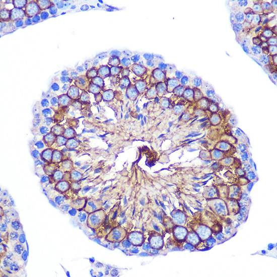
Immunohistochemistry analysis of paraffin-embedded Mouse testis using CD47 Rabbit pAb at dilution of 1:100 (40x lens). Microwave antigen retrieval performed with 0.01M PBS Buffer (pH 7.2) prior to IHC staining.

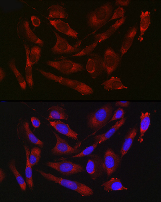
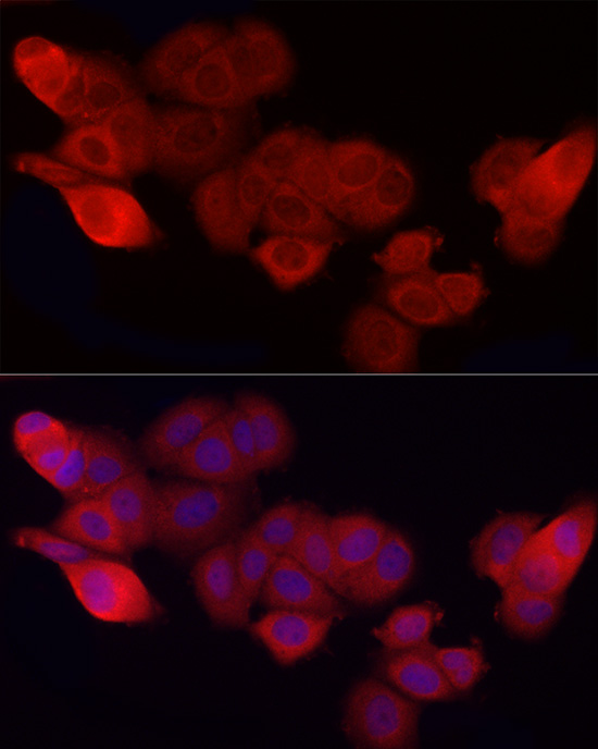
Immunofluorescence analysis of U2OS cells using CD47 Rabbit pAb at dilution of 1:50 (40x lens). Secondary antibody: Cy3-conjugated Goat anti-Rabbit IgG (H+L) at 1:500 dilution. Blue: DAPI for nuclear staining.

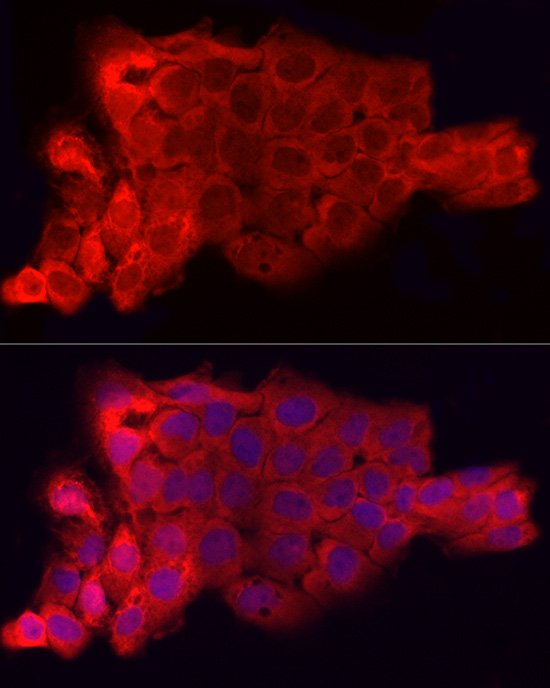
Immunofluorescence analysis of A431 cells using CD47 Rabbit pAb at dilution of 1:50 (40x lens). Secondary antibody: Cy3-conjugated Goat anti-Rabbit IgG (H+L) ) at 1:500 dilution. Blue: DAPI for nuclear staining.

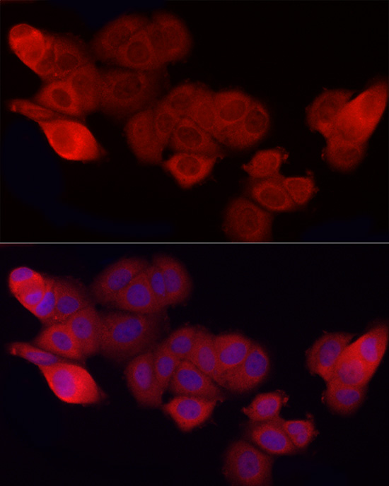
Immunofluorescence analysis of MCF7 cells using CD47 Rabbit pAb at dilution of 1:50 (40x lens). Secondary antibody: Cy3-conjugated Goat anti-Rabbit IgG (H+L) at 1:500 dilution. Blue: DAPI for nuclear staining.

No documents available.