CD86 Rabbit Polyclonal Antibody

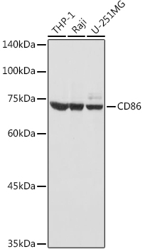
Western blot analysis of extracts of various cell lines, using CD86 antibody at 1:1000 dilution.  Secondary antibody: HRP Goat Anti-Rabbit IgG (H+L) at 1:10000 dilution.
Lysates/proteins: 25ug per lane.
Blocking buffer: 3% nonfat dry milk in TBST.
Detection: ECL West Pico Plus. 
Exposure time: 1s

Western blot analysis of extracts of various cell lines, using CD86 antibody at 1:1000 dilution. Secondary antibody: HRP Goat Anti-Rabbit IgG (H+L) at 1:10000 dilution. Lysates/proteins: 25ug per lane. Blocking buffer: 3% nonfat dry milk in TBST. Detection: ECL West Pico Plus. Exposure time: 1s

Catalog Number:AB-84415
Conjugate:Unconjugated
Size:100 ug
Concentration:1mg/ml
Host:Rabbit
Isotype:IgG
Clone:POLY
Immunogen:Synthesized peptide derived from human CD86
Reactivity:Human, Mouse, Rat
Applications:Western Blot: 1:500-2000 Immunofluorescence: 1:100-500 Immunocytochemistry: 1:100-500 ELISA: 1:5000-20000
Molecular Weight:60-80kD
Purification:The antibody was affinity-purified from rabbit antiserum by affinity-chromatography using epitope-specific immunogen.
Form:Liquid

Applications

Western Blot: 1:500-2000 Immunofluorescence: 1:100-500 Immunocytochemistry: 1:100-500 ELISA: 1:5000-20000

Immunogen

Synthesized peptide derived from human CD86
Target Background

This gene encodes a type I membrane protein that is a member of the immunoglobulin superfamily. This protein is expressed by antigen-presenting cells, and it is the ligand for two proteins at the cell surface of T cells, CD28 antigen and cytotoxic T-lymphocyte-associated protein 4. Binding of this protein with CD28 antigen is a costimulatory signal for activation of the T-cell. Binding of this protein with cytotoxic T-lymphocyte-associated protein 4 negatively regulates T-cell activation and diminishes the immune response. Alternative splicing results in several transcript variants encoding different isoforms.

Storage

Store at -20℃. Avoid repeated freeze-thaw cycles.

Buffer

Liquid in PBS containing 50% glycerol, 0.5% BSA and 0.02% sodium azide.

Western blot analysis of extracts of various cell lines, using CD86 antibody at 1:1000 dilution. Secondary antibody: HRP Goat Anti-Rabbit IgG (H+L) at 1:10000 dilution. Lysates/proteins: 25ug per lane. Blocking buffer: 3% nonfat dry milk in TBST. Detection: ECL West Pico Plus. Exposure time: 1s

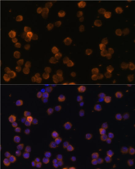
Immunofluorescence analysis of Raw264.7 cells using CD86 antibody at dilution of 1:100. Blue: DAPI for nuclear staining.

No documents available.