CK7 Mouse mAb

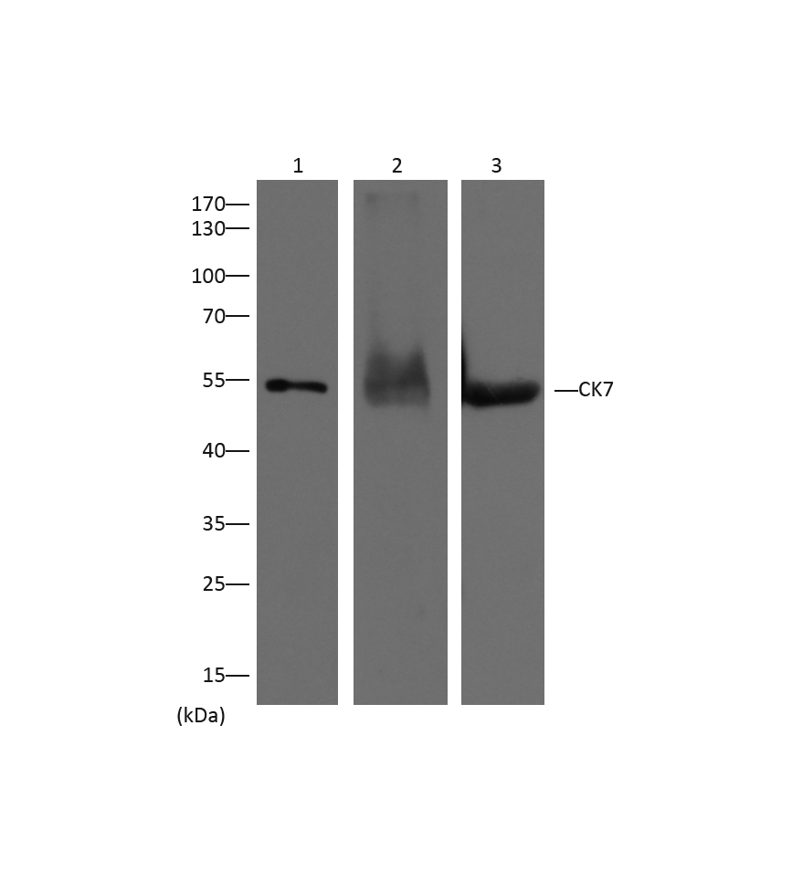
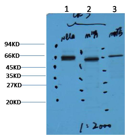
Western blot analysis of 1) Hela, 2)Mouse Kidney, 3) Mouse Brain with CK7 mAb diluted at 1:2,000.

Western blot analysis of 1) Hela, 2)Mouse Kidney, 3) Mouse Brain with CK7 mAb diluted at 1:2,000.

Catalog Number:ABE1054
Conjugate:unconjugated
Size:100 ug
Concentration:1mg/mL
Host:Mouse
Isotype:IgG1
Clone:Monoclonal
Immunogen:Synthetic Peptide
Reactivity:Human, Rat, Mouse
Applications:WB 1:1,000-2,000 IHC 1:200 IP 1:200
Molecular Weight:54kDa
Form:liquid

Cytokeratin 7 is a 54kD intermediate filament protein found in a variety of glandularepithelia. Cytokeratin 7 has been found in columnar and glandular epithelium of the lung, cervix, breast, bile ducts and larger collecting ducts of the kidney. It is present in the transitional epithelium of bladder as well as ovarian and lungepithelia, and occasionally staining of blood vessel cell walls, particularly endothelial cells, may be observed. However, Cytokeratin 7 is not expressed by epithelial cells of the gastrointestinal tract, colon or prostate. Keratin 7 is often co expressed with keratin 19.

Applications

WB 1:1,000-2,000 IHC 1:200 IP 1:200

Immunogen

Synthetic Peptide
Target Background

Cytokeratin 7 is a 54kD intermediate filament protein found in a variety of glandularepithelia. Cytokeratin 7 has been found in columnar and glandular epithelium of the lung, cervix, breast, bile ducts and larger collecting ducts of the kidney. It is present in the transitional epithelium of bladder as well as ovarian and lungepithelia, and occasionally staining of blood vessel cell walls, particularly endothelial cells, may be observed. However, Cytokeratin 7 is not expressed by epithelial cells of the gastrointestinal tract, colon or prostate. Keratin 7 is often co expressed with keratin 19.

Storage

PBS with 0.02% sodium azide and 50% glycerol pH 7.4. Store at -20°C. Avoid repeated freeze-thaw cycles.

Western blot analysis of 1) Hela, 2)Mouse Kidney, 3) Mouse Brain with CK7 mAb diluted at 1:2,000.

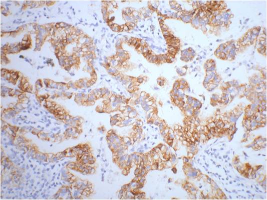
IHC staining of human lung cancer tissue with CK7 mouse mAb(12D7) diluted at 1:200.

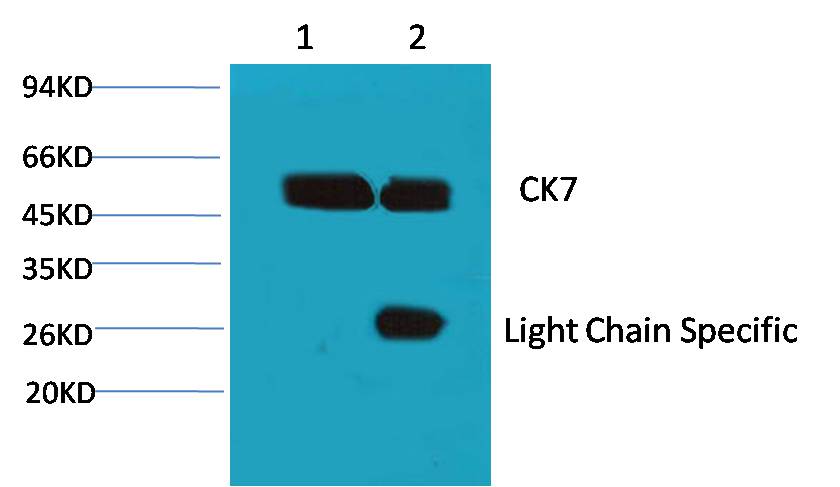
1、Input: Hela Cell Lysate 2、IP product: IP dilute 1:200 Western blot analysis: primary antibody : EM1054 1:1,000 Secondary antibody: Goat anti-Mouse IgG Light chain specific(S003), 1:5,000

No documents available.