Claudin-5 Rabbit Polyclonal Antibody

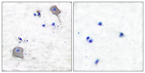
Immunohistochemistry analysis of paraffin-embedded human brain tissue, using Claudin 5 Antibody. The picture on the right is blocked with the synthesized peptide.

Immunohistochemistry analysis of paraffin-embedded human brain tissue, using Claudin 5 Antibody. The picture on the right is blocked with the synthesized peptide.

Catalog Number:ABN08911
Conjugate:Unconjugated
Size:100 ug
Concentration:1mg/ml
Host:Rabbit
Isotype:IgG
Clone:Polyclonal
Immunogen:The antiserum was produced against synthesized peptide derived from human Claudin 5. AA range:169-218
Reactivity:Human,Mouse,Rat
Applications:WB 1:500-1:2000,IHC 1:100-1:300,ICC/IF 1:100-1:300,ELISA 1:10000-1:20000
Molecular Weight:22kDa
Purification:Affinity purification
Form:Liquid
Synonyms:CLDN5; AWAL; TMVCF; Claudin-5; Transmembrane protein deleted in VCFS; TMDVCF

This gene encodes a member of the claudin family. Claudins are integral membrane proteins and components of tight junction strands. Tight junction strands serve as a physical barrier to prevent solutes and water from passing freely through the paracellular space between epithelial or endothelial cell sheets. Mutations in this gene have been found in patients with velocardiofacial syndrome. Alternatively spliced transcript variants encoding the same protein have been found for this gene. [provided by RefSeq, Aug 2008],function:Plays a major role in tight junction-specific obliteration of the intercellular space.,similarity:Belongs to the claudin family.,subunit:Directly interacts with TJP1/ZO-1, TJP2/ZO-2 and TJP3/ZO-3. Interacts with MPDZ.

Applications

WB 1:500-1:2000,IHC 1:100-1:300,ICC/IF 1:100-1:300,ELISA 1:10000-1:20000

Immunogen

The antiserum was produced against synthesized peptide derived from human Claudin 5. AA range:169-218

Target Background

This gene encodes a member of the claudin family. Claudins are integral membrane proteins and components of tight junction strands. Tight junction strands serve as a physical barrier to prevent solutes and water from passing freely through the paracellular space between epithelial or endothelial cell sheets. Mutations in this gene have been found in patients with velocardiofacial syndrome. Alternatively spliced transcript variants encoding the same protein have been found for this gene. [provided by RefSeq, Aug 2008],function:Plays a major role in tight junction-specific obliteration of the intercellular space.,similarity:Belongs to the claudin family.,subunit:Directly interacts with TJP1/ZO-1, TJP2/ZO-2 and TJP3/ZO-3. Interacts with MPDZ.

Synonyms:CLDN5; AWAL; TMVCF; Claudin-5; Transmembrane protein deleted in VCFS; TMDVCF

Storage

Aliquot and store at -20°C (valid for 12 months). Avoid freeze/thaw cycles.

Buffer

Liquid in PBS containing 50% glycerol, 0.5% protective protein and 0.02% New type preservative N.

Immunohistochemistry analysis of paraffin-embedded human brain tissue, using Claudin 5 Antibody. The picture on the right is blocked with the synthesized peptide.

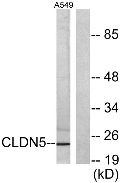
Western blot analysis of lysates from A549 cells, using Claudin 5 Antibody. The lane on the right is blocked with the synthesized peptide.

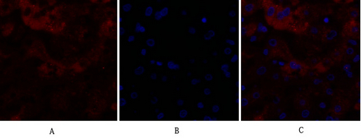
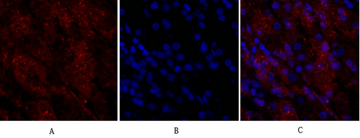
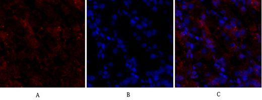
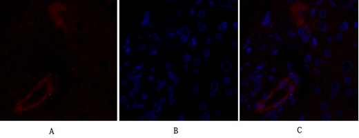
Immunofluorescence analysis of human-liver tissue. 1,Claudin-5 Polyclonal Antibody(red) was diluted at 1:200(4°C,overnight). 2, Cy3 labled Secondary antibody was diluted at 1:300(room temperature, 50min).3, Picture B: DAPI(blue) 10min. Picture A:Target. Picture B: DAPI. Picture C: merge of A+B

Immunofluorescence analysis of human-liver tissue. 1,Claudin-5 Polyclonal Antibody(red) was diluted at 1:200(4°C,overnight). 2, Cy3 labled Secondary antibody was diluted at 1:300(room temperature, 50min).3, Picture B: DAPI(blue) 10min. Picture A:Target. Picture B: DAPI. Picture C: merge of A+B

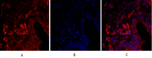
Immunofluorescence analysis of human-lung tissue. 1,Claudin-5 Polyclonal Antibody(red) was diluted at 1:200(4°C,overnight). 2, Cy3 labled Secondary antibody was diluted at 1:300(room temperature, 50min).3, Picture B: DAPI(blue) 10min. Picture A:Target. Picture B: DAPI. Picture C: merge of A+B

Immunofluorescence analysis of human-lung tissue. 1,Claudin-5 Polyclonal Antibody(red) was diluted at 1:200(4°C,overnight). 2, Cy3 labled Secondary antibody was diluted at 1:300(room temperature, 50min).3, Picture B: DAPI(blue) 10min. Picture A:Target. Picture B: DAPI. Picture C: merge of A+B

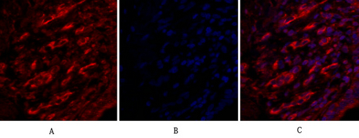
Immunofluorescence analysis of human-stomach tissue. 1,Claudin-5 Polyclonal Antibody(red) was diluted at 1:200(4°C,overnight). 2, Cy3 labled Secondary antibody was diluted at 1:300(room temperature, 50min).3, Picture B: DAPI(blue) 10min. Picture A:Target. Picture B: DAPI. Picture C: merge of A+B

Immunofluorescence analysis of human-stomach tissue. 1,Claudin-5 Polyclonal Antibody(red) was diluted at 1:200(4°C,overnight). 2, Cy3 labled Secondary antibody was diluted at 1:300(room temperature, 50min).3, Picture B: DAPI(blue) 10min. Picture A:Target. Picture B: DAPI. Picture C: merge of A+B

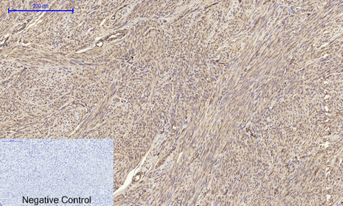
Immunohistochemical analysis of paraffin-embedded Human-uterus tissue. 1,Claudin-5 Polyclonal Antibody was diluted at 1:200(4°C,overnight). 2, Sodium citrate pH 6.0 was used for antibody retrieval(>98°C,20min). 3,Secondary antibody was diluted at 1:200(room tempeRature, 30min). Negative control was used by secondary antibody only.

No documents available.