Cleaved-PARP-1 (G215)

Western blot analysis of extracts from normal
(control) and PARP1 knockout (KO) HeLa cells,
using PARP antibody att 1:1000 dilution.
Secondary antibody: HRP Goat
Anti-Rabbit IgG (H+L) at 1:10000 dilution.
Lysates/proteins: 25ug per lane.
Blocking buffer: 3% nonfat dry milk in TBST.
Detection: ECL West Pico Plus.
Exposure time: 1s.

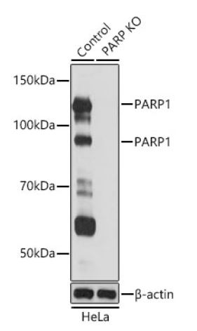
Western blot analysis of extracts from normal (control) and PARP1 knockout (KO) HeLa cells, using PARP antibody att 1:1000 dilution. Secondary antibody: HRP Goat Anti-Rabbit IgG (H+L) at 1:10000 dilution. Lysates/proteins: 25ug per lane. Blocking buffer: 3% nonfat dry milk in TBST. Detection: ECL West Pico Plus. Exposure time: 1s.

Catalog Number:AB-84347
Size:100ug
Concentration:1mg/ml
Host:Rb
Isotype:IgG
Clone:POLY
Immunogen:The antiserum was produced against synthesized peptide derived from human PARP. AA range:196-245
Reactivity:Hu, Ms
Applications:WB 1:1000 Immunohistochemistry 1 :50-1:200
Molecular Weight:89, 113kDa
Purification:The antibody was affinity-purified from rabbit antiserum by affinity chromatography using epitope-specific immunogen.
Form:Liquid

Applications

WB 1:1000 Immunohistochemistry 1 :50-1:200

Immunogen

The antiserum was produced against synthesized peptide derived from human PARP. AA range:196-245
Target Background

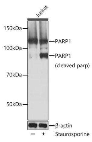
poly(ADP-ribose) polymerase 1(PARP1) Homo sapiens This gene encodes a chromatinassociated enzyme, poly(ADP-ribosyl)transferase, which modifies various nuclear proteins by poly(ADPribosyl) ation. The modification is dependent on DNA and is involved in the regulation of various important cellular processes such as differentiation, proliferation, and tumor transformation and also in the regulation of the molecular events involved in the recovery of cell from DNA damage. In addition, this enzyme may be the site of mutation in Fanconi anemia, and may participate in the pathophysiology of type I diabetes. The MW band of the cleaved form is recognized at 89kDa and the non-cleaved form is recognized at 113kDa.

Storage

20°C/1 year

Buffer

Liquid in PBS containing 50% glycerol, 0.5% BSA and 0.02% sodium azide.

Western blot analysis of extracts from normal (control) and PARP1 knockout (KO) HeLa cells, using PARP antibody att 1:1000 dilution. Secondary antibody: HRP Goat Anti-Rabbit IgG (H+L) at 1:10000 dilution. Lysates/proteins: 25ug per lane. Blocking buffer: 3% nonfat dry milk in TBST. Detection: ECL West Pico Plus. Exposure time: 1s.

Western blot analysis of extracts of Jurkat cells, using PARP1 antibody at 1:1000 dilution.Jurkat cells were treated by Staurosporine(1uM) at room temperature for 3 hours . Secondary antibody: HRP Goat Anti-Rabbit IgG (H+L) at 1:10000 dilution. Lysates/proteins: 25ug per lane. Blocking buffer: 3% nonfat dry milk in TBST. Detection: ECL West Pico Plus. Exposure time: 1s.

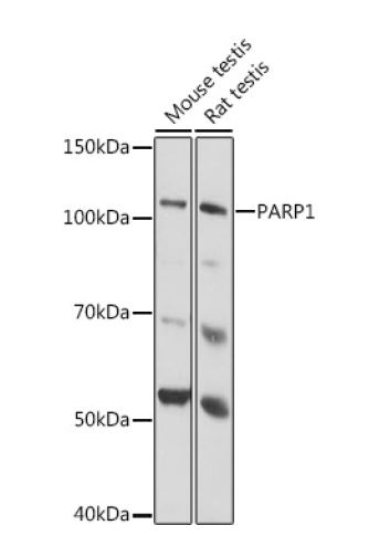
Western blot analysis of extracts of various cell lines, using PARP1 antibody at 1:1000 dilution. Secondary antibody: HRP Goat Anti-Rabbit IgG (H+L) at 1:10000 dilution. Lysates/proteins: 25ug per lane. Blocking buffer: 3% nonfat dry milk in TBST. Detection: ECL West Pico Plus. Exposure time: 1s.

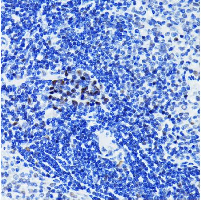
Immunohistochemistry of paraffin-embedded rat spleen using PARP1 antibody at dilution of 1:100 (40x lens).

No documents available.