Collagen Type I Rabbit Polyclonal Antibody
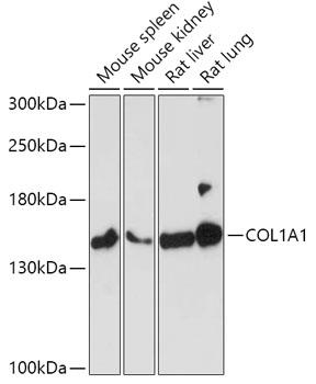
Western blot analysis of extracts of various cell lines, using COL1A1 antibody at 1:1000 dilution. Secondary antibody: HRP Goat Anti-Rabbit IgG (H+L) at 1:10000 dilution.
Lysates/proteins: 25ug per lane.
Blocking buffer: 3% nonfat dry milk in TBST.
Detection: ECL Basic Kit 
Exposure time: 3min.

Western blot analysis of extracts of various cell lines, using COL1A1 antibody at 1:1000 dilution. Secondary antibody: HRP Goat Anti-Rabbit IgG (H+L) at 1:10000 dilution. Lysates/proteins: 25ug per lane. Blocking buffer: 3% nonfat dry milk in TBST. Detection: ECL Basic Kit Exposure time: 3min.

Catalog Number:AB-82138
Conjugate:Unconjugated
Size:100 ug
Concentration:1mg/ml
Host:Rabbit
Isotype:IgG
Clone:POLY
Immunogen:A synthetic peptide corresponding to a sequence at the C-terminus of mouse Collagen I, identical to the related rat sequence, and different from the related human sequence by two amino acids.
Reactivity:Human, Mouse, Rat
Applications:Western Blot: 1:500 -1:2000Immunofluorescence: 1:50 - 1:200Immunocytochemistry: 1:50 - 1:200Immunohistochemistry: 1:50-1:200
Molecular Weight:140kDa/220kDa
Purification:Aff. Pur.
Form:Liquid
Synonyms:COL1A1; EDSC; OI1; OI2; OI3; OI4

Applications

Western Blot: 1:500 -1:2000Immunofluorescence: 1:50 - 1:200Immunocytochemistry: 1:50 - 1:200Immunohistochemistry: 1:50-1:200

Immunogen

A synthetic peptide corresponding to a sequence at the C-terminus of mouse Collagen I, identical to the related rat sequence, and different from the related human sequence by two amino acids.

Target Background

This gene encodes the pro-alpha1 chains of type I collagen whose triple helix comprises two alpha1 chains and one alpha2 chain. Type I is a fibril-forming collagen found in most connective tissues and is abundant in bone, cornea, dermis and tendon. Mutations in this gene are associated with osteogenesis imperfecta types I-IV, Ehlers- Danlos syndrome type VIIA, Ehlers-Danlos syndrome Classical type, Caffey Disease and idiopathic osteoporosis. Reciprocal translocations between chromosomes 17 and 22, where this gene and the gene for platelet-derived growth factor beta are located, are associated with a particular type of skin tumor called dermatofibrosarcoma protuberans, resulting from unregulated expression of the growth factor. Two transcripts, resulting from the use of alternate polyadenylation signals, have been identified for this gene.

Synonyms:COL1A1; EDSC; OI1; OI2; OI3; OI4

Storage

Store at -20℃. Avoid freeze / thaw cycles.

Buffer

Each vial contains 4mg Trehalose, 0.9mg NaCl and 0.2mg Na2HPO4.

Western blot analysis of extracts of various cell lines, using COL1A1 antibody at 1:1000 dilution. Secondary antibody: HRP Goat Anti-Rabbit IgG (H+L) at 1:10000 dilution. Lysates/proteins: 25ug per lane. Blocking buffer: 3% nonfat dry milk in TBST. Detection: ECL Basic Kit Exposure time: 3min.

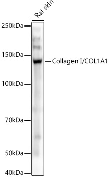
Western blot analysis of Rat skin, using CollagenI/COL1A1 antibody at 1:800 dilution. Secondary antibody: HRP Goat Anti-Rabbit IgG (H+L) at 1:10000 dilution. Lysates/proteins: 25ug per lane. Blocking buffer: 3% nonfat dry milk in TBST. Detection: ECL West Pico Plus. Exposure time: 3s.

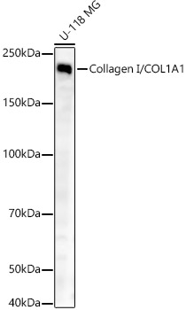
Western blot analysis of U-118 MG, using CollagenI/COL1A1 antibody at 1:800 dilution. Secondary antibody: HRP Goat Anti-Rabbit IgG (H+L) at 1:10000 dilution. Lysates/proteins: 25ug per lane. Blocking buffer: 3% nonfat dry milk in TBST. Detection: ECL West Pico Plus. Exposure time: 10s.

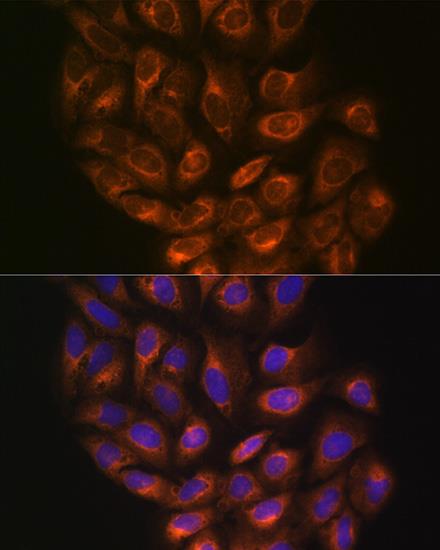
Immunofluorescence analysis of NIH/3T3 cells using Collagen I/COL1A1 Rabbit pAb (A1352) at dilution of 1:50 (40x lens). Blue: DAPI for nuclear staining.

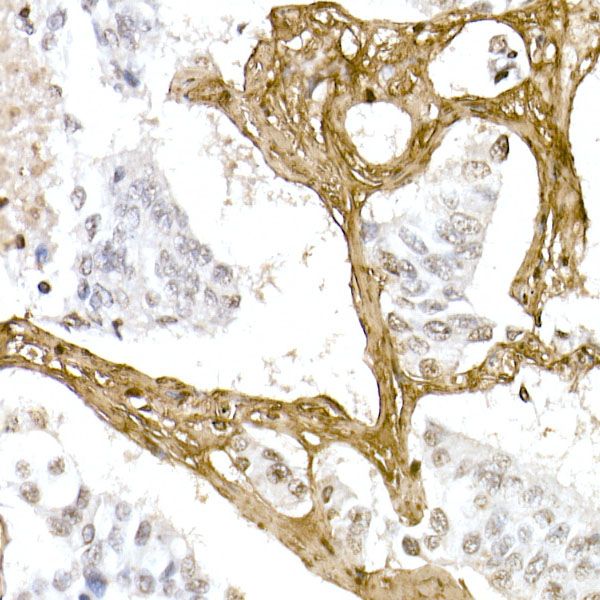
Immunohistochemistry of paraffinembedded human lung cancer using Collagen I/COL1A1 Rabbit pAb at dilution of 1:100 (40x lens).Perform microwave antigen retrieval with 10 mM PBS buffer pH 7.2 before commencing with IHC staining protocol.

No documents available.