
Collagen III alpha 1/COL3A1 Rabbit Polyclonal Antibody
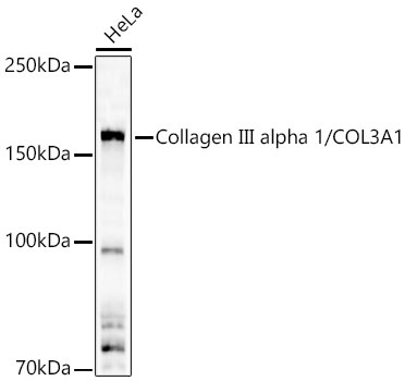
Western blot analysis of lysates from HeLa cells using Collagen III alpha 1/COL3A1 Rabbit pAb at 1:10000 dilution.
Secondary antibody:HRP Goat Anti-Rabbit IgG (H+L) at 1:10000 dilution.
Lysates/proteins: 25 μg per lane.
Blocking buffer: 3% nonfat dry milk in TBST.
Detection:ECL West Pico Plus.
Exposuretime:90s.

Western blot analysis of lysates from HeLa cells using Collagen III alpha 1/COL3A1 Rabbit pAb at 1:10000 dilution. Secondary antibody:HRP Goat Anti-Rabbit IgG (H+L) at 1:10000 dilution. Lysates/proteins: 25 μg per lane. Blocking buffer: 3% nonfat dry milk in TBST. Detection:ECL West Pico Plus. Exposuretime:90s.

Catalog Number:AB-82620
Conjugate:Unconjugated
Size:100 ug
Concentration:1mg/ml
Host:Rabbit
Isotype:IgG
Clone:POLY
Immunogen:Recombinant fusion protein containing a sequence corresponding to amino acids 1201-1301 of human Collagen III alpha 1/COL3A1.
Reactivity:Hu , Ms
Applications:Western Blot: 1:2000 - 1:10000Immunofluorescence: 1:50 - 1:200Immunocytochemistry. 1:50 - 1:200
Molecular Weight:150kDa
Purification:Affinity purification
Form:Liquid
Synonyms:EDS4A; EDSVASC; PMGEDSV; Collagen III alpha 1/COL3A1

Applications

Western Blot: 1:2000 - 1:10000Immunofluorescence: 1:50 - 1:200Immunocytochemistry. 1:50 - 1:200

Immunogen

Recombinant fusion protein containing a sequence corresponding to amino acids 1201-1301 of human Collagen III alpha 1/COL3A1.

Target Background

This gene encodes the pro-alpha1 chains of type III collagen, a fibrillar collagen that is found in extensible connective tissues such as skin, lung, uterus, intestine and the vascular system, frequently in association with type I collagen. Mutations in this gene are associated with Ehlers-Danlos syndrome type IV, and with aortic and arterial aneurysms.

Synonyms:EDS4A; EDSVASC; PMGEDSV; Collagen III alpha 1/COL3A1

Storage

Store at -20℃. Avoid freeze / thaw cycles.

Buffer

PBS with 0.02% sodium azide,50% glycerol,pH7.3.

Western blot analysis of lysates from HeLa cells using Collagen III alpha 1/COL3A1 Rabbit pAb at 1:10000 dilution. Secondary antibody:HRP Goat Anti-Rabbit IgG (H+L) at 1:10000 dilution. Lysates/proteins: 25 μg per lane. Blocking buffer: 3% nonfat dry milk in TBST. Detection:ECL West Pico Plus. Exposuretime:90s.

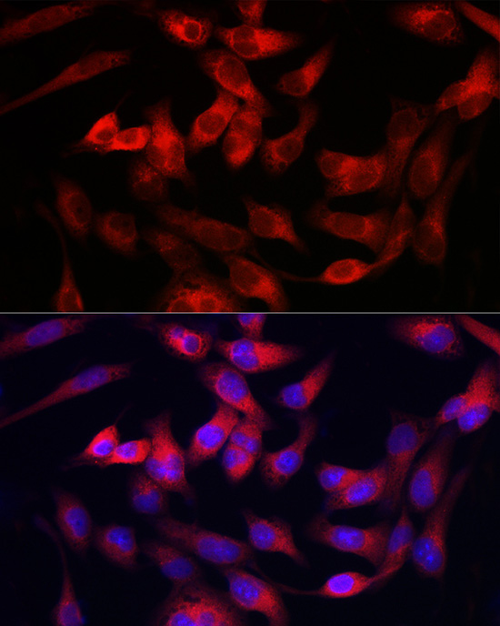
Immunofluorescence analysis of RD cells using Collagen III alpha 1/COL3A1 Rabbit pAb at dilution of 1:100. Secondary antibody: Cy3 Goat Anti-Rabbit IgG (H+L) (AS007) at 1:500 dilution. Blue: DAPI for nuclear staining.

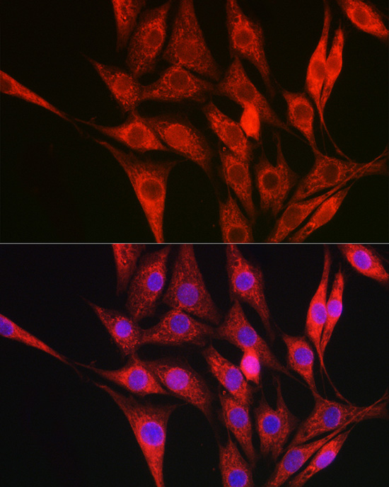
Immunofluorescence analysis of NIH/3T3 cells using Collagen III alpha 1/COL3A1 Rabbit pAb at dilution of 1:100. Secondary antibody: Cy3 Goat Anti-Rabbit IgG (H+L) at 1:500 dilution. Blue: DAPI for nuclear staining.

No documents available.