
Collagen Type IV

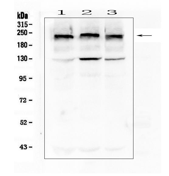
Western blot analysis of Collagen IV using anti-
Collagen IV antibody ( Electrophoresis was performed on a 8% SDS-PAGE gel at 70V (Stacking gel) / 90V (Resolving gel) for 2-3 hours. The sample well of each lane was loaded with 50ug of sample under
reducing conditions.
Lane 1: human HEK293T whole cell lysate,
Lane 2: human Hela whole cell lysate,
Lane 3: human A549 whole cell lysate.
After Electrophoresis, proteins were transferred to a Nitrocellulose membrane at 150mA for 50-90 minutes. Blocked the membrane with 5% Non-fat Milk/ TBS for 1.5 hour at RT. The membrane was incubated with mouse anti-Collagen IV antigen affinity purified monoclonal antibody at 0.5 μg/mL overnight at 4°C, then washed with TBS-0.1%Tween 3 times with 5 minutes each and probed with a goat anti-mouse IgG-HRP secondary antibody at a dilution of 1:10000 for 1.5 hour at RT. The signal is developed using an Enhanced Chemiluminescent detection (ECL1) with Tanon 5200 system. A specific band was detected for Collagen IV at approximately 220KD. The expected band size for Collagen IV is at 161KD.

Western blot analysis of Collagen IV using anti- Collagen IV antibody ( Electrophoresis was performed on a 8% SDS-PAGE gel at 70V (Stacking gel) / 90V (Resolving gel) for 2-3 hours. The sample well of each lane was loaded with 50ug of sample under reducing conditions. Lane 1: human HEK293T whole cell lysate, Lane 2: human Hela whole cell lysate, Lane 3: human A549 whole cell lysate. After Electrophoresis, proteins were transferred to a Nitrocellulose membrane at 150mA for 50-90 minutes. Blocked the membrane with 5% Non-fat Milk/ TBS for 1.5 hour at RT. The membrane was incubated with mouse anti-Collagen IV antigen affinity purified monoclonal antibody at 0.5 μg/mL overnight at 4°C, then washed with TBS-0.1%Tween 3 times with 5 minutes each and probed with a goat anti-mouse IgG-HRP secondary antibody at a dilution of 1:10000 for 1.5 hour at RT. The signal is developed using an Enhanced Chemiluminescent detection (ECL1) with Tanon 5200 system. A specific band was detected for Collagen IV at approximately 220KD. The expected band size for Collagen IV is at 161KD.

Catalog Number:MABX-91945
Conjugate:Unconjugated
Size:100 ug
Concentration:1mg/ml
Host:Ms
Isotype:IgG1
Clone:3G3
Immunogen:E.coli-derived human Collagen IV recombinant protein (Position: G1445-T1669). Human Collagen IV shares 97% amino acid (aa) sequence identity with mouse Collagen IV.
Reactivity:Human
Applications:Western Blot: 1:2000-1:1000 Immunohistochemistry(Paraffin-embedded Section): 1:1000 Immunofluorescence: 1:250-1:100
Purification:Aff. Pur.
Form:Liquid

Applications

Western Blot: 1:2000-1:1000 Immunohistochemistry(Paraffin-embedded Section): 1:1000 Immunofluorescence: 1:250-1:100

Immunogen

E.coli-derived human Collagen IV recombinant protein (Position: G1445-T1669). Human Collagen IV shares 97% amino acid (aa) sequence identity with mouse Collagen IV.

Target Background

COL4A1, also known as ICH or Collagen alpha-1(IV), is a protein that in humans is encoded by the COL4A1 gene. It is mapped to 13q34. This gene encodes the major type IV alpha collagen chain of basement membranes. Like the other members of the type IV collagen gene family, this gene is organized in a head-to-head conformation with another type IV collagen gene so that each gene pair shares a common promoter. COL4A1 binds to alpha-1/beta-1 integrin and inhibits migration, proliferation, and tube formation by endothelial cells. It is also a potential therapeutic candidate for targeting tumor angiogenesis.

Storage

At -20°C for one year. It can also be aliquotted and stored frozen at -20°C for a longer time. Avoid repeated freezing and thawing.

Buffer

Each vial contains 4mg Trehalose, 0.9mg NaCl, 0.2mg Na2HPO4, 0.05mg NaN3.

Western blot analysis of Collagen IV using anti- Collagen IV antibody ( Electrophoresis was performed on a 8% SDS-PAGE gel at 70V (Stacking gel) / 90V (Resolving gel) for 2-3 hours. The sample well of each lane was loaded with 50ug of sample under reducing conditions. Lane 1: human HEK293T whole cell lysate, Lane 2: human Hela whole cell lysate, Lane 3: human A549 whole cell lysate. After Electrophoresis, proteins were transferred to a Nitrocellulose membrane at 150mA for 50-90 minutes. Blocked the membrane with 5% Non-fat Milk/ TBS for 1.5 hour at RT. The membrane was incubated with mouse anti-Collagen IV antigen affinity purified monoclonal antibody at 0.5 μg/mL overnight at 4°C, then washed with TBS-0.1%Tween 3 times with 5 minutes each and probed with a goat anti-mouse IgG-HRP secondary antibody at a dilution of 1:10000 for 1.5 hour at RT. The signal is developed using an Enhanced Chemiluminescent detection (ECL1) with Tanon 5200 system. A specific band was detected for Collagen IV at approximately 220KD. The expected band size for Collagen IV is at 161KD.

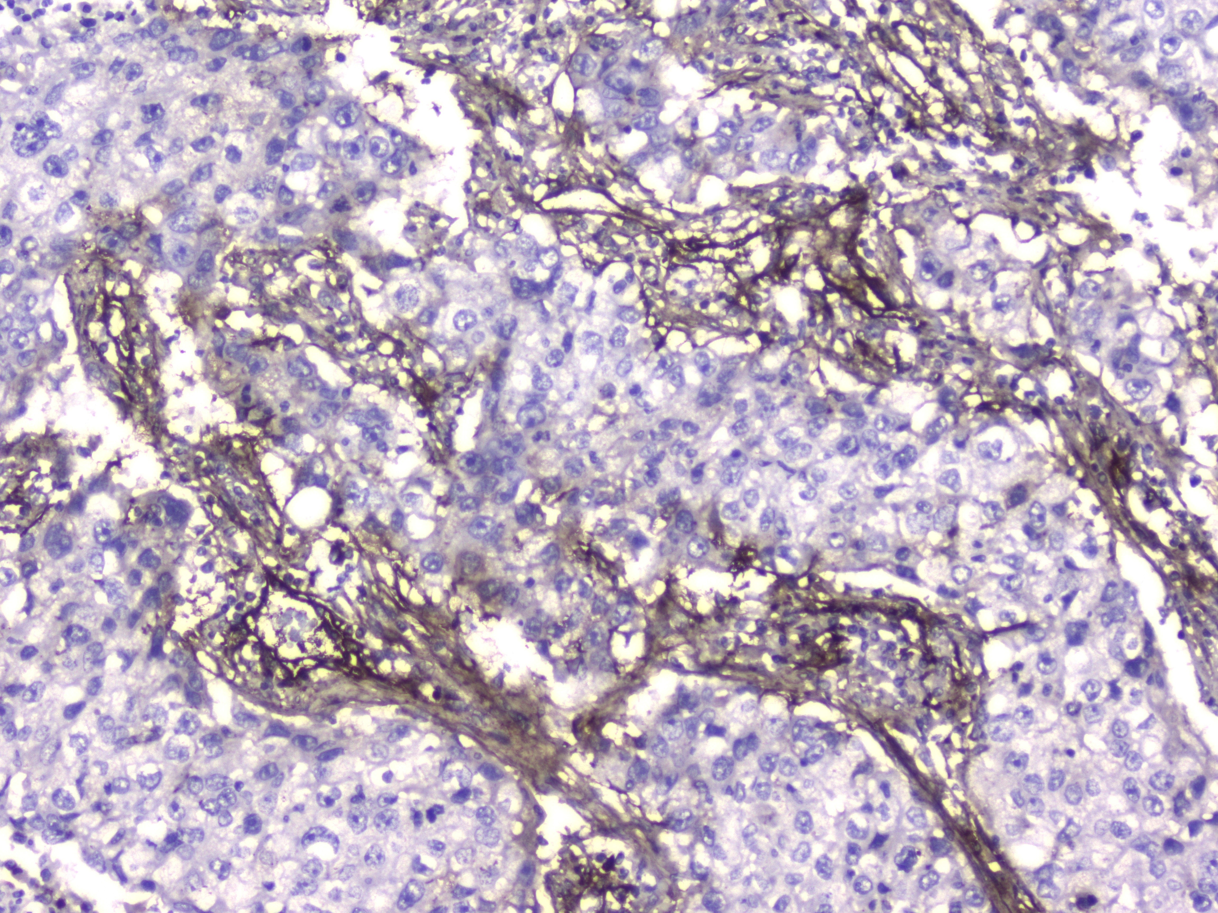
IHC analysis of Collagen IV using anti-Collagen IV antibody . Collagen IV was detected in paraffin-embedded section of human lung cancer tissue. Heat mediated antigen retrieval was performed in citrate buffer (pH6, epitope retrieval solution) for 20 mins. The tissue section was blocked with 10% goat serum. The tissue section was then incubated with 2μg/ml mouse anti- Collagen IV Antibody overnight at 4°C. Biotinylated goat anti-mouse IgG was used as secondary antibody and incubated for 30 minutes at 37°C. The tissue section was developed using Strepavidin-Biotin-Complex (SABC) with DAB as the chromogen.

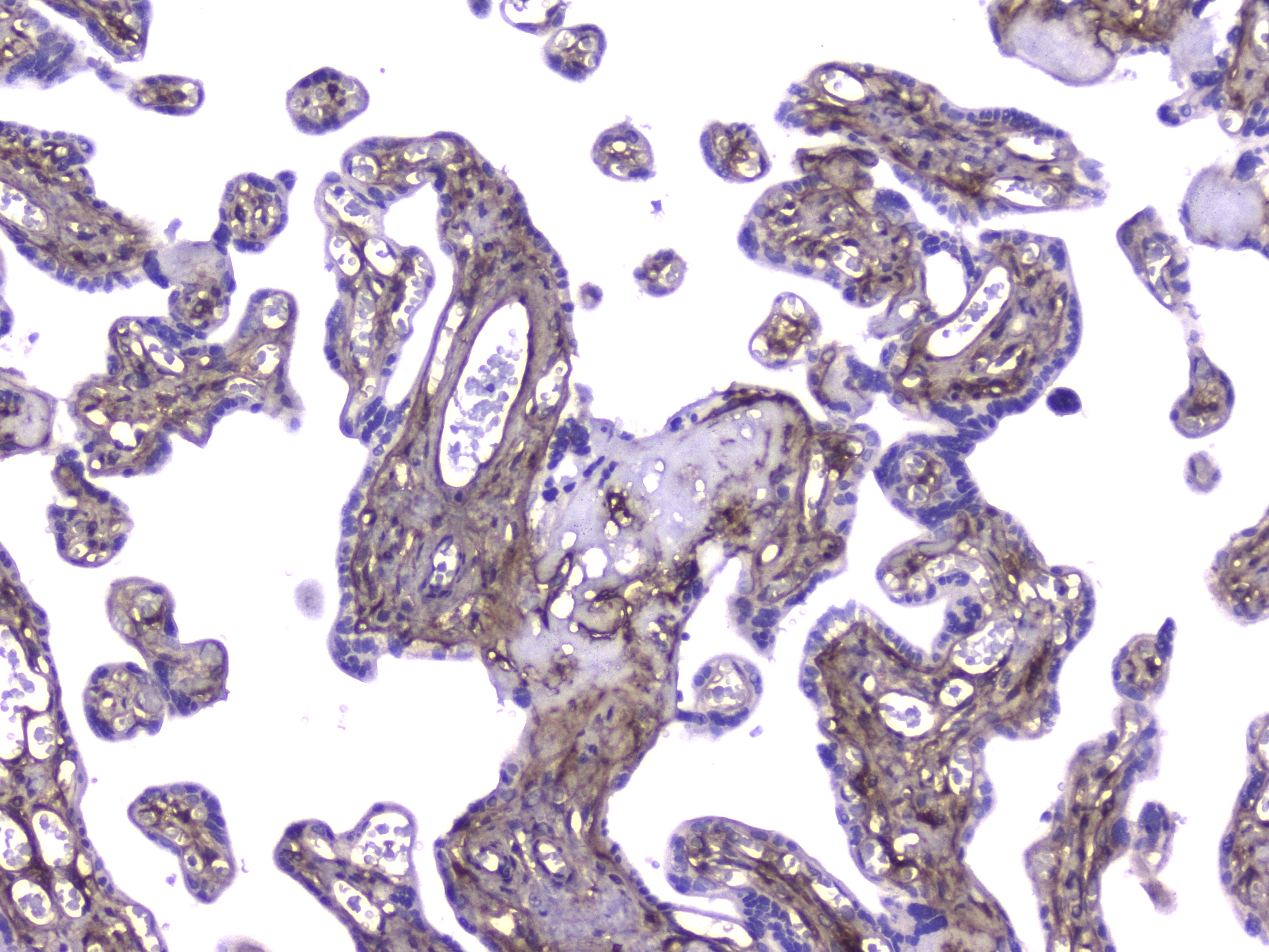
IHC analysis of Collagen IV using anti-Collagen IV antibody Collagen IV was detected in paraffin-embedded section of human placenta tissue. Heat mediated antigen retrieval was performed in citrate buffer (pH6, epitope retrieval solution) for 20 mins. The tissue section was blocked with 10% goat serum. The tissue section was then incubated with 2μg/ml mouse anti- Collagen IV Antibody overnight at 4°C. Biotinylated goat anti-mouse IgG was used as secondary antibody and ,incubated for 30 minutes at 37°C. The tissue section was developed using Strepavidin-Biotin-Complex (SABC) with DAB as the chromogen.

No documents available.