COX 2

tfistern blot analysis of extracts of various cell
lines, using Cox2 antibody at 1:1000 dilution.
Secondary antibody: HRP Goat Anti-Rabbit IgG
(H+L) at 1:10000 dilution. Lysates/proteins:
25ug per lane. Blocking buffer: 3% nonfat dry milk
in TBST.
Detection: ECL West Pico Plus.
Exposure time: 3s.

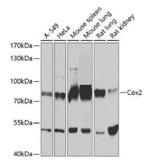
tfistern blot analysis of extracts of various cell lines, using Cox2 antibody at 1:1000 dilution. Secondary antibody: HRP Goat Anti-Rabbit IgG (H+L) at 1:10000 dilution. Lysates/proteins: 25ug per lane. Blocking buffer: 3% nonfat dry milk in TBST. Detection: ECL West Pico Plus. Exposure time: 3s.

Catalog Number:MAB-94444
Size:100 ug
Concentration:1mg/ml
Host:Rb
Isotype:IgG
Clone:D5H5
Reactivity:Hu, Ms, Rt
Applications:WB 1:1000-1:2000, IHC-P 1:100, IF-IP 1:100, Unmasking buffer: Citrate
Molecular Weight:74 kDa
Purification:Monoclonal antibody is produced by immunizing animals with a synthetic peptide corresponding to residues surrounding His108 of human Cox2 protein.
Form:Liquid

Applications

WB 1:1000-1:2000, IHC-P 1:100, IF-IP 1:100, Unmasking buffer: Citrate
Target Background

Cyclooxygenase1 (Cox1) and cyclooxygenase2 (Cox2), family members with 60% homology in humans, catalyze prostaglandin production from arachidonic acid (1,2). While Cox1 expression is constitutive in most tissues, Cox2 expression is induced by lipopolysaccharide (LPS) and peptidoglycan (PGN) (3). PGN activates Ras, leading to phosphorylation of Raf at Ser338 and Erk1/2 at Tyr204. The activation of MAP kinase signaling results in subsequent activation of IKKα/β, phosphorylation of IκBα at Ser32/36, and NF-κB activation. Finally, activation of the transcription factor NF-κB is responsible for the induction of Cox2 expression (4). Investigators have shown that LPS and PGN induce the clinical manifestations of arthritis and bacterial infections, such as inflammation, fever, and septic shock (5). Research studies have indicated that Cox1 and Cox2 may also play a role in the neuropathology of Alzheimer’s disease by potentiating γ-secretase activity and β-amyloid generation (6).Cox2 (D5H5) XP® Rabbit mAb recognizes endogenous levels of total Cox2 protein.

Storage

Store at -20°C. Avoid freeze/thaw cycles

Buffer

PBS with 0.02% sodium azide, 50%glycerol, pH7.3.

tfistern blot analysis of extracts of various cell lines, using Cox2 antibody at 1:1000 dilution. Secondary antibody: HRP Goat Anti-Rabbit IgG (H+L) at 1:10000 dilution. Lysates/proteins: 25ug per lane. Blocking buffer: 3% nonfat dry milk in TBST. Detection: ECL West Pico Plus. Exposure time: 3s.

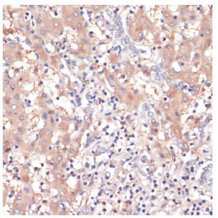
Immunohistochemistry of paraffinembedded human liver cancer using Cox2 antibody at dilution of 1:200 (40x lens).

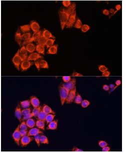
Immunofluorescence analysis of HeLa cells using Cox2 antibody at dilution of 1:100. Blue: DAPI for nuclear staining.

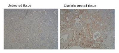
Application Immunohistochemistry(IHC) Sample RAW264.7,Mouse kidney Dilution 1:200 Incubatition time 12h Temperature 4 ℃ Exposure unknow Observed Band unknow kDa Remarks Immunohistochemistry of paraffin-embedded mouse kidney tissue using ptgs2(cox-2 )antibody at dilution of 1:100 (x100 lens).

No documents available.