CRALBP Rabbit Monoclonal Antibody

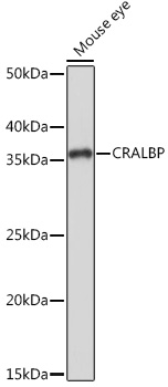
Western blot analysis of extracts of Mouse eye, using CRALBP Rabbit mAb at 1:1000 dilution.
Secondary antibody: HRP Goat Anti-Rabbit IgG (H+L)  at 1:10000 dilution.
Lysates/proteins: 25μg per lane.
Blocking buffer: 3% nonfat dry milk in TBST.
Detection: ECL West Pico Plus.
Exposure time: 1s.

Western blot analysis of extracts of Mouse eye, using CRALBP Rabbit mAb at 1:1000 dilution. Secondary antibody: HRP Goat Anti-Rabbit IgG (H+L) at 1:10000 dilution. Lysates/proteins: 25μg per lane. Blocking buffer: 3% nonfat dry milk in TBST. Detection: ECL West Pico Plus. Exposure time: 1s.

Catalog Number:MAB-94793
Conjugate:Unconjugated
Size:100 ug
Concentration:1mg/ml
Host:Rabbit
Isotype:IgG
Clone:ARC1502
Immunogen:A synthetic peptide corresponding to a sequence within amino acids 1-100 of human CRALBP
Reactivity:Human, Mouse, Rat
Applications:Western Blot: 1:500 - 1:2000 Immunohistochemistry(paraffin-embedded tissues): 1:50 - 1:200 Immunofluorescence: 1:50 - 1:200 Immunocytochemistry: 1:50 - 1:200
Molecular Weight:36kDa
Purification:Affinity purification
Form:Liquid
Synonyms:CRALBP

Applications

Western Blot: 1:500 - 1:2000 Immunohistochemistry(paraffin-embedded tissues): 1:50 - 1:200 Immunofluorescence: 1:50 - 1:200 Immunocytochemistry: 1:50 - 1:200

Immunogen

A synthetic peptide corresponding to a sequence within amino acids 1-100 of human CRALBP
Target Background

The protein encoded by this gene is a 36-kD water-soluble protein which carries 11-cisretinaldehyde or 11-cis-retinal as physiologic ligands. It may be a functional component of the visual cycle. Mutations of this gene have been associated with severe rod-cone dystrophy, Bothnia dystrophy (nonsyndromic autosomal recessive retinitis pigmentosa) and retinitis punctata albescens.

Synonyms:CRALBP

Storage

Store at -20°C. Avoid repeated freeze-thaw cycles.

Buffer

36kDa

Western blot analysis of extracts of Mouse eye, using CRALBP Rabbit mAb at 1:1000 dilution. Secondary antibody: HRP Goat Anti-Rabbit IgG (H+L) at 1:10000 dilution. Lysates/proteins: 25μg per lane. Blocking buffer: 3% nonfat dry milk in TBST. Detection: ECL West Pico Plus. Exposure time: 1s.

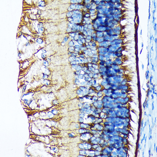
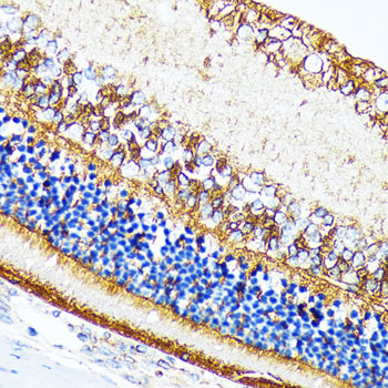
Immunohistochemistry analysis of paraffin-embedded rat retina using CRALBP Rabbit mAb at dilution of n1:100 (40x lens).Perform microwave antigen retrieval with 10 mM Tris/EDTA buffer pH 9.0 before commencing with IHC staining protocol.

Immunohistochemistry analysis of paraffin-embedded mouse retina using CRALBP Rabbit mAb at dilution of 1:100 (40x lens).Perform microwave antigen retrieval with 10 mM Tris/EDTA buffer pH 9.0 before commencing with IHC staining protocol.

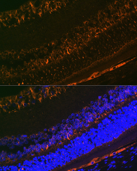
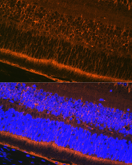
Immunofluorescence analysis of rat eye using CRALBP Rabbit mAb at dilution of 1:100 (40x lens). Blue: DAPI for nuclear staining.

Immunofluorescence analysis of mouse eye using CRALBP Rabbit mAb at dilution of 1:100 (40x lens). Blue: DAPI for nuclear staining.

No documents available.