CXCR2 Rabbit Polyclonal Antibody

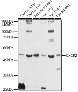
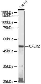
Western blot analysis of various lysates using CXCR2 Rabbit pAb at 1:1000 dilution.
Secondary antibody: HRP Goat Anti-Rabbit IgG (H+L) at 1:10000 dilution.
Lysates/proteins: 25μg per lane.
Blocking buffer: 3% nonfat dry milk in TBST.
Detection: ECL West Pico Plus. 
Exposure time: 10s.

Western blot analysis of various lysates using CXCR2 Rabbit pAb at 1:1000 dilution. Secondary antibody: HRP Goat Anti-Rabbit IgG (H+L) at 1:10000 dilution. Lysates/proteins: 25μg per lane. Blocking buffer: 3% nonfat dry milk in TBST. Detection: ECL West Pico Plus. Exposure time: 10s.

Catalog Number:AB-90154
Conjugate:Unconjugated
Size:100 ug
Concentration:1mg/ml
Host:Rabbit
Isotype:IgG
Clone:POLY
Immunogen:A synthetic peptide corresponding to a sequence within amino acids 260-359 of human CXCR2
Reactivity:Human,Mouse,Rat
Applications:Western Blot: 1:500 - 1:1000 Immunohistochemistry (paraffin-embedded tissues): 1:50 - 1:200 Immunofluorescence: 1:50 - 1:200 Immunocytochemistry: 1:50 - 1:200
Molecular Weight:46kDa
Purification:Affinity purification
Form:Liquid
Synonyms:CD182; IL8R2; IL8RA; IL8RB; CMKAR2; WHIMS2; CDw128b; CXCR2

Applications

Western Blot: 1:500 - 1:1000 Immunohistochemistry (paraffin-embedded tissues): 1:50 - 1:200 Immunofluorescence: 1:50 - 1:200 Immunocytochemistry: 1:50 - 1:200

Immunogen

A synthetic peptide corresponding to a sequence within amino acids 260-359 of human CXCR2
Target Background

The protein encoded by this gene is a member of the G-protein-coupled receptor family. This protein is a receptor for interleukin 8 (IL8). It binds to IL8 with high affinity, and transduces the signal through a G-protein activated second messenger system. This receptor also binds to chemokine (C-X-C motif) ligand 1 (CXCL1/MGSA), a protein with melanoma growth stimulating activity, and has been shown to be a major component required for serum-dependent melanoma cell growth. This receptor mediates neutrophil migration to sites of inflammation. The angiogenic effects of IL8 in intestinal microvascular endothelial cells are found to be mediated by this receptor. Knockout studies in mice suggested that this receptor controls the positioning of oligodendrocyte precursors in developing spinal cord by arresting their migration. This gene, IL8RA, a gene encoding another high affinity IL8 receptor, as well as IL8RBP, a pseudogene of IL8RB, form a gene cluster in a region mapped to chromosome 2q33-q36. Alternatively spliced variants, encoding the same protein, have been identified.

Synonyms:CD182; IL8R2; IL8RA; IL8RB; CMKAR2; WHIMS2; CDw128b; CXCR2

Storage

Store at -20℃. Avoid freeze / thaw cycles.

Buffer

PBS with 0.01% thimerosal,50% glycerol,pH7.3.

Western blot analysis of various lysates using CXCR2 Rabbit pAb at 1:1000 dilution. Secondary antibody: HRP Goat Anti-Rabbit IgG (H+L) at 1:10000 dilution. Lysates/proteins: 25μg per lane. Blocking buffer: 3% nonfat dry milk in TBST. Detection: ECL West Pico Plus. Exposure time: 10s.

Western blot analysis of lysates from THP-1 cells, using CXCR2 Rabbit pAb at 1:1000 dilution. Secondary antibody: HRP Goat Anti-Rabbit IgG (H+L) at 1:10000 dilution. Lysates/proteins: 25μg per lane. Blocking buffer: 3% nonfat dry milk in TBST. Detection: ECL West Pico Plus. Exposure time: 30s.

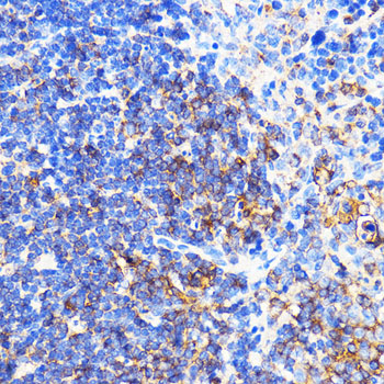
Immunohistochemistry analysis of paraffin-embedded Mouse spleen using CXCR2 Rabbit pAb at dilution of 1:100 (40x lens).Perform microwave antigen retrieval with 10 mM PBS buffer pH 7.2 before commencing with IHC staining protocol.

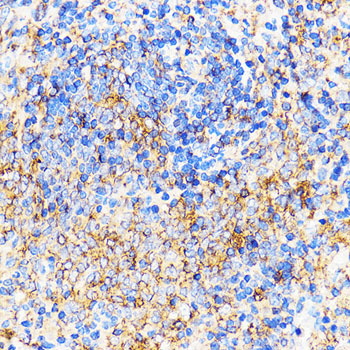
Immunohistochemistry analysis of paraffin-embedded Rat spleen using CXCR2 Rabbit pAb at dilution of 1:100 (40x lens).Perform microwave antigen retrieval with 10 mM PBS buffer pH 7.2 befor commencing with IHC staining protocol.

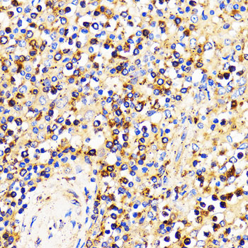
Immunohistochemistry analysis of paraffin-embedded Human spleen using CXCR2 Rabbit pAb at dilution of 1:100 (40x lens).Perform microwave antigen retrieval with 10 mM PBS buffer pH 7.2 before commencing with IHC staining protocol.

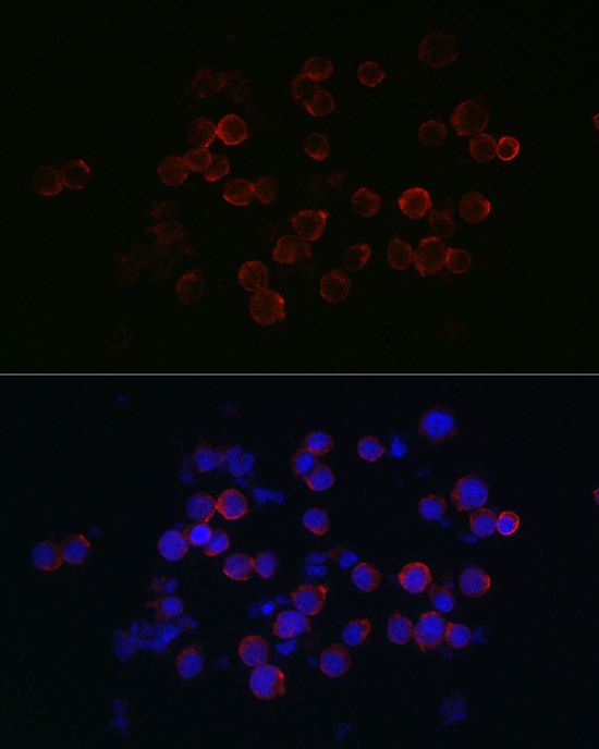
Immunofluorescence analysis of Jurkat cells using CXCR2 Rabbit pAb at dilution of 1:200 (40xlens). Secondary antibody: Cy3 Goat Anti-Rabbit IgG (H+L) at 1:500 dilution. Blue: DAPI for nuclear staining.

No documents available.