CXCR4 Rabbit Polyclonal Antibody

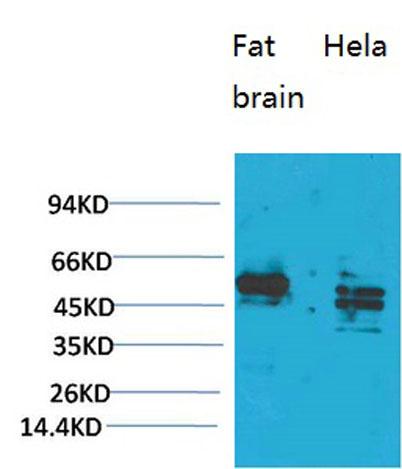
Western blot analysis of 1) Rat Brain Tissue, 2)Hela, with CXCR4 Rabbit pAb diluted at 1:1,000.

Western blot analysis of 1) Rat Brain Tissue, 2)Hela, with CXCR4 Rabbit pAb diluted at 1:1,000.

Catalog Number:AB-83811
Conjugate:Unconjugated
Size:100 ug
Concentration:1mg/ml
Host:Rabbit
Isotype:IgG
Clone:POLY
Immunogen:Synthetic Peptide of CXCR4 AA range: 203-253
Reactivity:Human,Mouse,Rat
Applications:Western Blot: 1:1000-2000 Immunohistochemistry: 1:100-200
Molecular Weight:50kD
Purification:The antibody was affinity-purified from rabbit antiserum by affinity-chromatography using epitope-specific immunogen.
Form:Liquid
Synonyms:CXCR4; C-X-C chemokine receptor type 4; CXC-R4; CXCR-4; FB22; Fusin; HM89; LCR1; Leukocyte-derived seven transmembrane domain receptor; LESTR; NPYRL; Stromal cell-derived factor 1 receptor; SDF-1 receptor; CD antigen CD184

Applications

Western Blot: 1:1000-2000 Immunohistochemistry: 1:100-200

Immunogen

Synthetic Peptide of CXCR4 AA range: 203-253
Target Background

C-X-C motif chemokine receptor 4(CXCR4) Homo sapiens This gene encodes a CXC chemokine receptor specific for stromal cell-derived factor-1. The protein has 7 transmembrane regions and is located on the cell surface. It acts with the CD4 protein to support HIV entry into cells and is also highly expressed in breast cancer cells. Mutations in this gene have been associated with WHIM (warts, hypogammaglobulinemia, infections, and myelokathexis) syndrome. Alternate transcriptional splice variants, encoding different isoforms, have been characterized.

Synonyms:CXCR4; C-X-C chemokine receptor type 4; CXC-R4; CXCR-4; FB22; Fusin; HM89; LCR1; Leukocyte-derived seven transmembrane domain receptor; LESTR; NPYRL; Stromal cell-derived factor 1 receptor; SDF-1 receptor; CD antigen CD184

Storage

Store at -20°C. Avoid repeated freeze-thaw cycles.

Buffer

Liquid in PBS containing 50% glycerol, 0.5% BSA and 0.02% sodium azide.

Western blot analysis of 1) Rat Brain Tissue, 2)Hela, with CXCR4 Rabbit pAb diluted at 1:1,000.

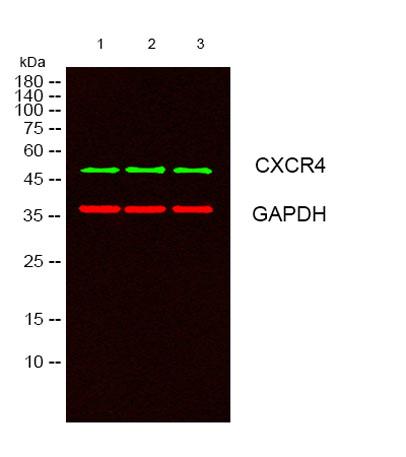
Western blot analysis of lysates from 1) Rat Brain Tissue, 2)Hela, 3) HEK293 cells, (Green) primary antibody was diluted at 1:1000, 4°over night, secondary antibody Dk x Rb was diluted at 1:10000, 37° 1hour. (Red) GAPDH Monoclonal Antibody(2B8) antibody was diluted at 1:5000 as loading control, 4° overnight,secondary antibody (Gt x Ms was diluted at 1:10000, 37° 1hour.

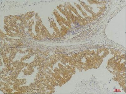
Immunohistochemical analysis of paraffin-embedded Human Breast Carcinoma using CXCR4Rabbit pAb diluted at 1:200.

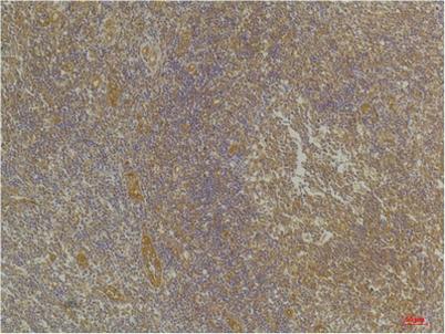
Immunohistochemical analysis of paraffin-embedded Human Tonsil Tissue using CXCR4Rabbit pAb diluted at 1:200.

No documents available.