DDC Rabbit Rabbit Polyclonal Antibody

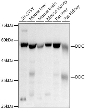
Western blot analysis of various lysates using DDC Rabbit pAb at 1:500 dilution.
Secondary antibody: HRP-conjugated Goat anti-Rabbit IgG (H+L)  at 1:10000 dilution.
Lysates/proteins: 25μg per lane.
Blocking buffer: 3% nonfat dry milk in TBST.
Detection: ECL West Pico Plus.  
Exposure time: 3s.

Western blot analysis of various lysates using DDC Rabbit pAb at 1:500 dilution. Secondary antibody: HRP-conjugated Goat anti-Rabbit IgG (H+L) at 1:10000 dilution. Lysates/proteins: 25μg per lane. Blocking buffer: 3% nonfat dry milk in TBST. Detection: ECL West Pico Plus. Exposure time: 3s.

Catalog Number:AB3828
Conjugate:Unconjugated
Size:100 ug
Concentration:1mg/ml
Host:Rabbit
Isotype:IgG
Clone:POLY
Immunogen:Recombinant protein.This information is considered to be commercially sensitive.
Reactivity:Human,Mouse,Rat
Applications:WB 1:200 - 1:800 IF-P 1:50 - 1:200 IHC-P 1:50 - 1:200 ELISA Recommended starting concentration is 1 μg/mL. Please optimize the concentration based on your specific assay requirements.
Molecular Weight:37kDa/54kDa
Purification:Affinity purification
Form:liquid
Synonyms:AADC; DDC

The encoded protein catalyzes the decarboxylation of L-3,4-dihydroxyphenylalanine (DOPA) to dopamine, L-5-hydroxytryptophan to serotonin and L-tryptophan to tryptamine. Defects in this gene are the cause of aromatic L-amino-acid decarboxylase deficiency (AADCD). AADCD deficiency is an inborn error in neurotransmitter metabolism that leads to combined serotonin and catecholamine deficiency. Multiple alternatively spliced transcript variants encoding different isoforms have been identified for this gene.

Applications

WB 1:200 - 1:800 IF-P 1:50 - 1:200 IHC-P 1:50 - 1:200 ELISA Recommended starting concentration is 1 μg/mL. Please optimize the concentration based on your specific assay requirements.

Immunogen

Recombinant protein.This information is considered to be commercially sensitive.
Target Background

The encoded protein catalyzes the decarboxylation of L-3,4-dihydroxyphenylalanine (DOPA) to dopamine, L-5-hydroxytryptophan to serotonin and L-tryptophan to tryptamine. Defects in this gene are the cause of aromatic L-amino-acid decarboxylase deficiency (AADCD). AADCD deficiency is an inborn error in neurotransmitter metabolism that leads to combined serotonin and catecholamine deficiency. Multiple alternatively spliced transcript variants encoding different isoforms have been identified for this gene.

Synonyms:AADC; DDC

Storage

Store at -20℃. Avoid freeze / thaw cycles.

Buffer

PBS containing 50% glycerol, preserved with proclin300 or sodium azide (as specified on the Certificate of Analysis),pH7.3.

Western blot analysis of various lysates using DDC Rabbit pAb at 1:500 dilution. Secondary antibody: HRP-conjugated Goat anti-Rabbit IgG (H+L) at 1:10000 dilution. Lysates/proteins: 25μg per lane. Blocking buffer: 3% nonfat dry milk in TBST. Detection: ECL West Pico Plus. Exposure time: 3s.

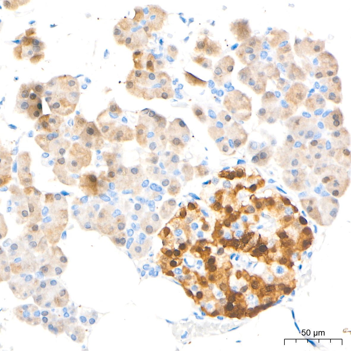
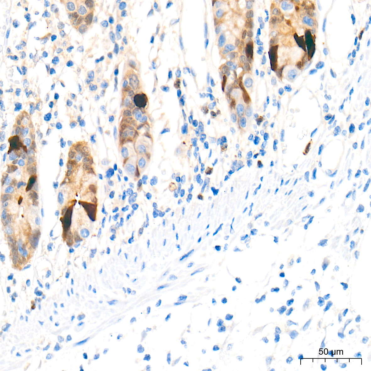
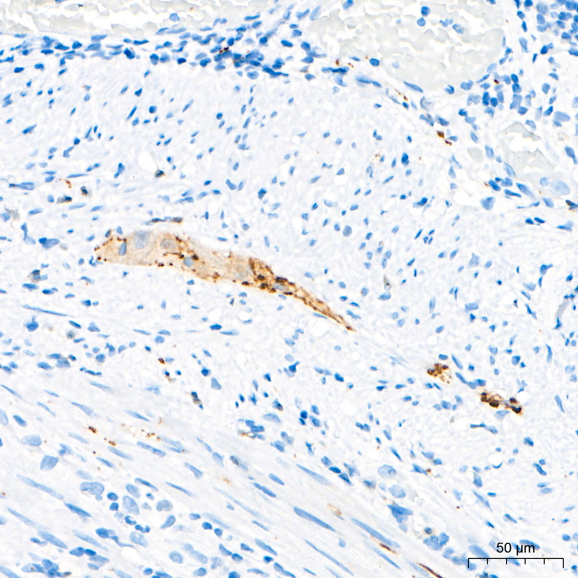
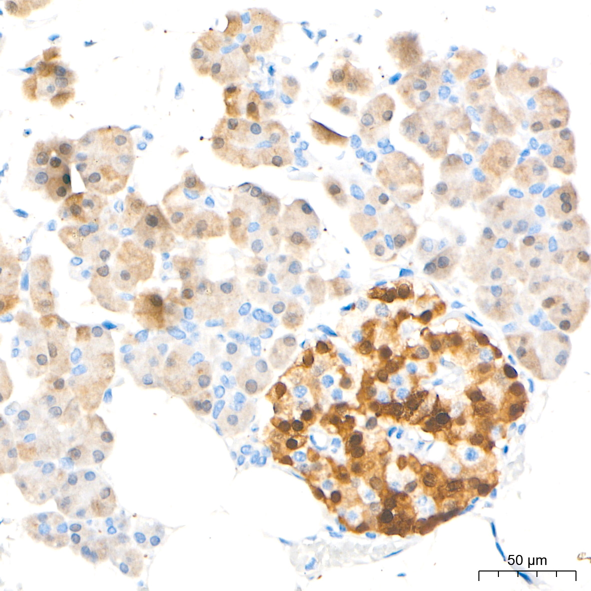
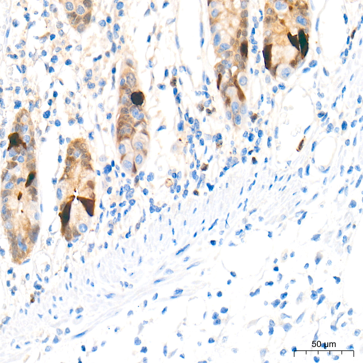
Immunohistochemistry analysis of paraffin-embedded tissue using DDC Rabbit pAb at a dilution of 1:800 (40x lens). High pressure antigen retrieval performed with 0.01M Tris-EDTA Buffer (pH 9.0) prior to IHC staining.

Immunohistochemistry analysis of paraffin-embedded tissue using DDC Rabbit pAb at a dilution of 1:800 (40x lens). High pressure antigen retrieval performed with 0.01M Tris-EDTA Buffer (pH 9.0) prior to IHC staining.

Immunohistochemistry analysis of paraffin-embedded Human colon tissue using DDC Rabbit pAb at a dilution of 1:1000 (40x lens). High pressure antigen retrieval performed with 0.01M Tris-EDTA Buffer (pH 9.0) prior to IHC staining.

Immunohistochemistry analysis of paraffin-embedded Human appendix tissue using DDC Rabbit pAb at a dilution of 1:1000 (40x lens). High pressure antigen retrieval performed with 0.01M Tris-EDTA Buffer (pH 9.0) prior to IHC staining.

Immunohistochemistry analysis of paraffin-embedded Human pancreas tissue using DDC Rabbit pAb at a dilution of 1:1000 (40x lens). High pressure antigen retrieval performed with 0.01M Tris-EDTA Buffer (pH 9.0) prior to IHC staining.

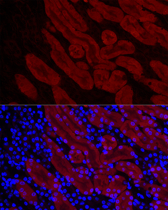
Immunofluorescence analysis of paraffin-embedded mouse kidney using DDC Rabbit pAb at dilution of 1:50 (40x lens). Secondary antibody: Cy3-conjugated Goat anti-Rabbit IgG (H+L) at 1:500 dilution. Blue: DAPI for nuclear staining.

No documents available.