E-cadherin Rabbit Polyclonal Antibody
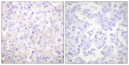
Immunohistochemistry analysis of paraffin-embedded human breast carcinoma tissue, using E-Cadherin Antibody. The picture on the right is Blocked with the synthesized peptide.
| Catalog Number: | ABN10278 |
| Conjugate: | Unconjugated |
| Size: | 100 ug |
| Concentration: | 1 mg/ml |
| Host: | Rabbit |
| Isotype: | IgG |
| Clone: | Polyclonal |
| Immunogen: | The antiserum was produced against synthesized peptide derived from human Cadherin. range: 1-50 |
| Reactivity: | Human,Mouse,Rat |
| Applications: | WB 1:500-1:1000; IHC 1:100-1:200; ICC/IF 1:100-1:200; ELISA 1:10000-1:20000 |
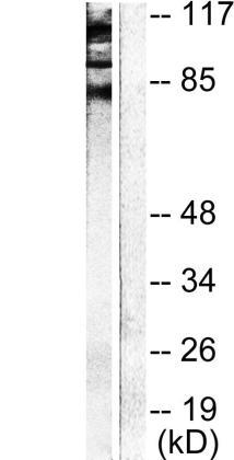
| Molecular Weight: | Calculated MW: 97 kDa |
| Purification: | Affinity purification |
| Form: | Liquid |
| Synonyms: | CDH1; CDHE; UVO; Cadherin-1; CAM120/80; Epithelial cadherin; E-cadherin |
Anti-E-cadherin Antibody detects endogenous levels of total E-cadherin protein. CDH1 (cadherin 1) codes for a transmembrane glycoprotein E-cadherin that is located on human chromosome 16q22.1. It is a tumor suppressor gene, which is also known as Cdh1p/Hct1p, Fizzy-related, Ste9, or Srw1.
Applications
WB 1:500-1:1000; IHC 1:100-1:200; ICC/IF 1:100-1:200; ELISA 1:10000-1:20000
Immunogen
The antiserum was produced against synthesized peptide derived from human Cadherin. range: 1-50