EGFR / HER1 Rabbit Polyclonal Antibody

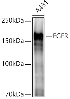
Western blot analysis of lysates from A431 cells, using EGFR Rabbit pAb at 1:2000 dilution.
Secondary antibody: HRP Goat Anti-Rabbit IgG (H+L) at 1:10000 dilution.
Lysates/proteins: 25ug per lane.
Blocking buffer: 3% nonfat dry milk in TBST.
Detection: ECL West Pico Plus. 
Exposure time: 3s.

Western blot analysis of lysates from A431 cells, using EGFR Rabbit pAb at 1:2000 dilution. Secondary antibody: HRP Goat Anti-Rabbit IgG (H+L) at 1:10000 dilution. Lysates/proteins: 25ug per lane. Blocking buffer: 3% nonfat dry milk in TBST. Detection: ECL West Pico Plus. Exposure time: 3s.

Catalog Number:AB-81864
Conjugate:Unconjugated
Size:100 ug
Concentration:1mg/ml
Host:Rabbit
Isotype:IgG
Clone:POLY
Immunogen:Recombinant fusion protein containing a sequence corresponding to amino acids 1021-1210 of human EGFR
Reactivity:Human,Mouse,Rat
Applications:Western Blot: 1:1000 - 1:5000 mmunohistochemistry(paraffin-embedded tissues) 1:50 - 1:200 Immunofluorescence: 1:100 - 1:500 Immuncytochemistry: 1:100 - 1:500 IImmunoprecipitation: 0.5μg-4μg antibody for 200μg-400μg extracts of whole cells
Molecular Weight:175kDa
Purification:Aff. Pur.
Form:Liquid
Synonyms:ERBB; ERRP; HER1; mENA; ERBB1; PIG61; NISBD2; EGFR

Applications

Western Blot: 1:1000 - 1:5000 mmunohistochemistry(paraffin-embedded tissues) 1:50 - 1:200 Immunofluorescence: 1:100 - 1:500 Immuncytochemistry: 1:100 - 1:500 IImmunoprecipitation: 0.5μg-4μg antibody for 200μg-400μg extracts of whole cells

Immunogen

Recombinant fusion protein containing a sequence corresponding to amino acids 1021-1210 of human EGFR

Target Background

The protein encoded by this gene is a transmembrane glycoprotein that is a member of the protein kinase superfamily. This protein is a receptor for members of the epidermal growth factor family. EGFR is a cell surface protein that binds to epidermal growth factor. Binding of the protein to a ligand induces receptor dimerization and tyrosine autophosphorylation and leads to cell proliferation. Mutations in this gene are associated with lung cancer.

Synonyms:ERBB; ERRP; HER1; mENA; ERBB1; PIG61; NISBD2; EGFR

Storage

Store at -20°C, and avoid repeat freeze-thaw cycles.

Buffer

PBS with 0.01% thimerosal,50% glycerol,pH7.3.

Western blot analysis of lysates from A431 cells, using EGFR Rabbit pAb at 1:2000 dilution. Secondary antibody: HRP Goat Anti-Rabbit IgG (H+L) at 1:10000 dilution. Lysates/proteins: 25ug per lane. Blocking buffer: 3% nonfat dry milk in TBST. Detection: ECL West Pico Plus. Exposure time: 3s.

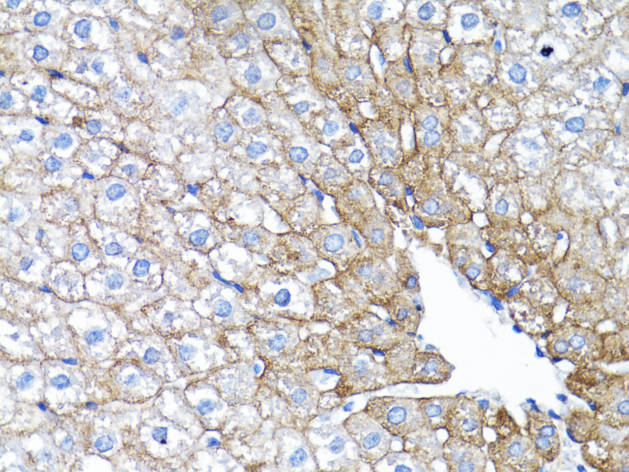
Immunohistochemistry analysis of paraffinembedded Rat liver using EGFR Rabbit pAb at dilution of 1:200 (40xlens).Perform microwave antigen retrieval with 10 mM PBS buffer pH 7.2 before commencing with IHC staining protocol.

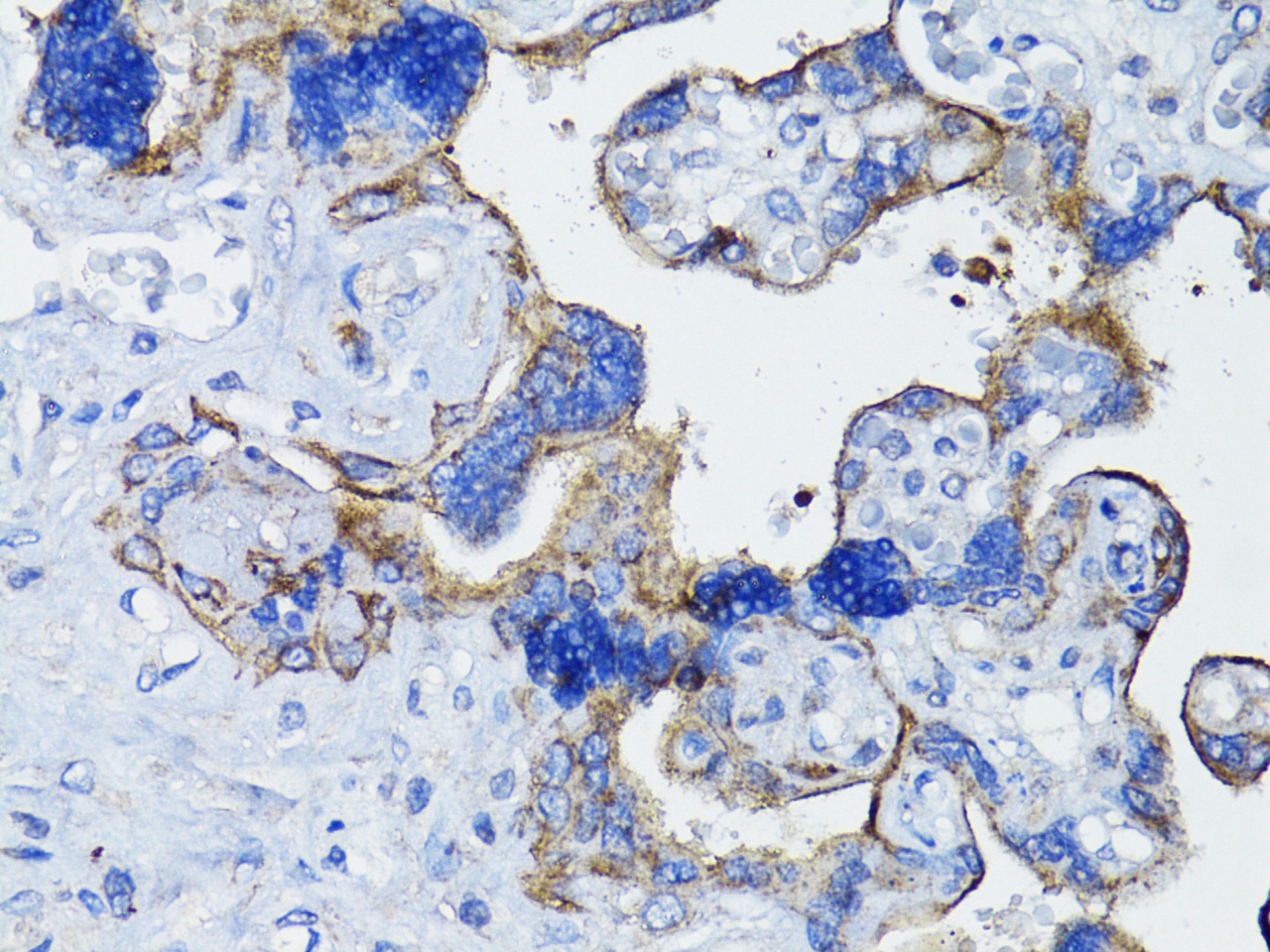
Immunohistochemistry analysis of paraffinembedded Human placenta using EGFR Rabbit pAb at dilution of 1:200 (40x lens).Perform microwave antigen retrieval with 10 mM PBS buffer pH 7.2 before commencing with IHC staining protocol.

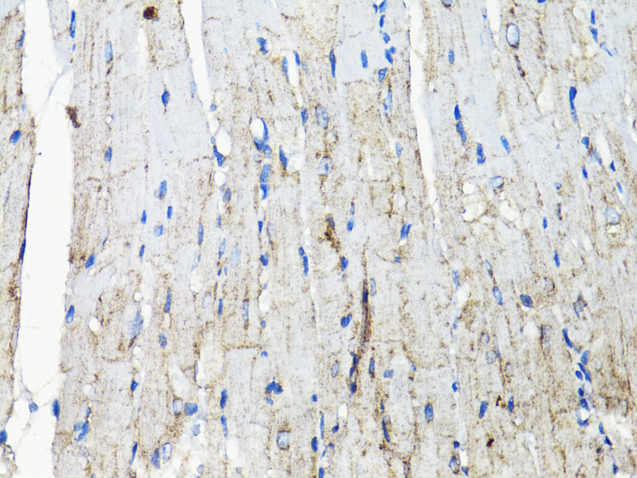
Immunohistochemistry analysis of paraffinembedded Mouse heart using EGFR Rabbit pAb at dilution of 1:200 (40x lens).Perform microwave antigen retrieval with 10 mM PBS buffer pH 7.2 before commencing with IHC staining protocol.

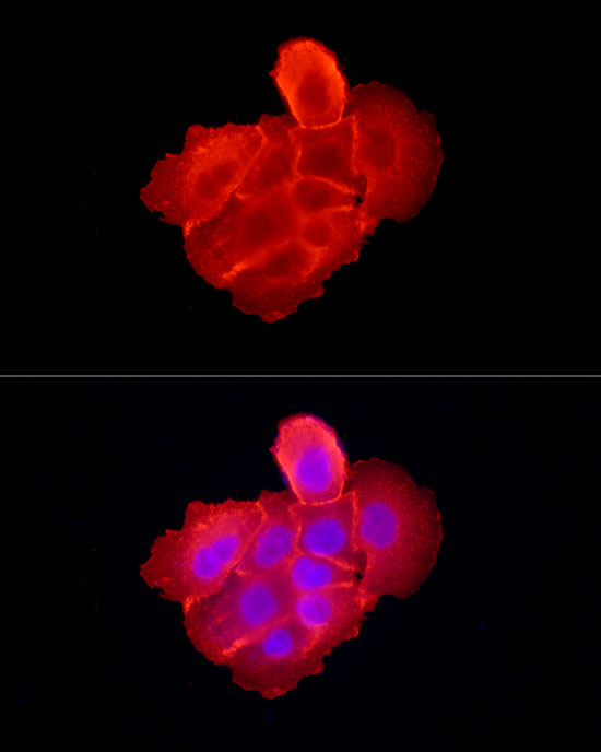
Immunofluorescence analysis of A-431 cells using EGFR Rabbit pAb at dilution of 1:300 (40x lens). Secondary antibody: Cy3 Goat Anti-Rabbit IgG (H+L) at 1:500 dilution. Blue: DAPI for nuclear staining.

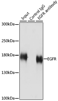
Immunoprecipitation analysis of 200 μg extracts of HeLa cells using 3 μg EGFR antibody. Western blot was performed from the immunoprecipitate using EGFR antibody at a dilution of 1:1000.

No documents available.