EPHA2

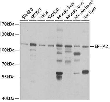
Western blot analysis of extracts of various cell lines, using EPHA2 antibody 1:1000 dilution.
Secondary antibody: HRP Goat Anti- Rabbit IgG (H+L)at 1:10000
dilution.
Lysates/proteins: 25ug per lane.
Blocking buffer: 3% nonfat dry milk in TBST.
Detection: ECL West Pico Plus
Exposure time: 10s.

Western blot analysis of extracts of various cell lines, using EPHA2 antibody 1:1000 dilution. Secondary antibody: HRP Goat Anti- Rabbit IgG (H+L)at 1:10000 dilution. Lysates/proteins: 25ug per lane. Blocking buffer: 3% nonfat dry milk in TBST. Detection: ECL West Pico Plus Exposure time: 10s.

Catalog Number:MAB-94587
Size:200 ug
Concentration:1mg/ml
Host:Rb
Isotype:IgG
Clone:A2/EPHA2
Immunogen:Recombinant fusion protein containing a sequence corresponding to amino acids 847-976 of human EPHA2
Reactivity:Hu, Ms, Rt
Applications:Western Blot: 1:500-1:2000 Immunohistochemistry (paraffin-embedded tissues): 1:50-1:200
Purification:Aff. Pur.
Form:Liquid

Applications

Western Blot: 1:500-1:2000 Immunohistochemistry (paraffin-embedded tissues): 1:50-1:200

Immunogen

Recombinant fusion protein containing a sequence corresponding to amino acids 847-976 of human EPHA2

Target Background

EPHA2(ephrin type-A receptor 2) also known as ECK, is a protein that in humans is encoded by the EPHA2 gene. This gene belongs to the ephrin receptor subfamily of the protein-tyrosine kinase family. Receptors in the EPH subfamily typically have a single kinase domain and an extracellular region containing a Cys-rich domain and 2 fibronectin type III repeats. By somatic cell hybrid analysis and fluorescence in situ hybridization, the EPHA2 gene is mapped to chromosome 1p36.1. By screening a HeLa cell cDNA library with degenerate oligonucleotides based on highly conserved regions of receptor protein-tyrosine kinases, Lindberg and Hunter isolated cDNAs encoding EPHA2, which they called ECK. EPHA2 was readily detectable in human lens fiber cells using immunoblot and immunohistochemistry. EGFR and EPHA2 mediated HCV entry by regulating CD81 -claudin-1(CLDN1) coreceptor associations and viral glycoprotein-dependent membrane fusion.

Storage

At -20°C for one year. Avoid repeated freezing and thawing.

Buffer

PBS with 0.02% sodium azide,50% glycerol,pH7.3.

Western blot analysis of extracts of various cell lines, using EPHA2 antibody 1:1000 dilution. Secondary antibody: HRP Goat Anti- Rabbit IgG (H+L)at 1:10000 dilution. Lysates/proteins: 25ug per lane. Blocking buffer: 3% nonfat dry milk in TBST. Detection: ECL West Pico Plus Exposure time: 10s.

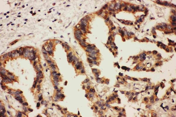
Anti- EPHA2 Antibody IHC(P) IHC(P): Human Lung Cancer Tissue

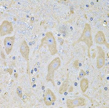
Immunohistochemistry of paraffin embedded rat brain using EPHA2 Antibody at dilution of 1:100 (40x lens).

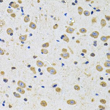
Immunohistochemistry of paraffin embedded mouse brain using EPHA2 Antibody at dilution of 1:100 (40x lens).

No documents available.