
FCGR2A

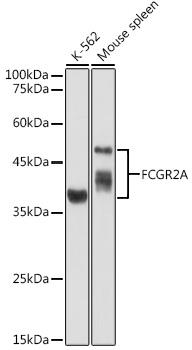
Western blot analysis of extracts of various cell lines, 
using FCGR2A antibody 1:1000dilution.
Secondary antibody: HRP Goat Anti-Rabbit IgG
 (H+L) at 1:10000 dilution.
Lysates/proteins: 25ug per lane.
Blocking buffer: 3% nonfat dry milk in TBST.
Detection: ECL West Pico Plus. 
Exposure time: 90s.

Western blot analysis of extracts of various cell lines, using FCGR2A antibody 1:1000dilution. Secondary antibody: HRP Goat Anti-Rabbit IgG (H+L) at 1:10000 dilution. Lysates/proteins: 25ug per lane. Blocking buffer: 3% nonfat dry milk in TBST. Detection: ECL West Pico Plus. Exposure time: 90s.

Catalog Number:AB-84343
Size:100 ug
Concentration:1mg/ml
Host:Rb
Isotype:IgG
Clone:POLY
Immunogen:Recombinant fusion protein containing a sequence corresponding to amino acids 34-316 of human FCGR2A (NP_067674.2).
Reactivity:Hu, Ms
Applications:Western Blot: 1:500 - 1:2000 Immunofluorescence: 1:50 - 1:200
Molecular Weight:40-50kDa
Purification:Aff. Pur.
Form:Liquid
Synonyms:FCGR2A;CD32;CD32A;CDw32;FCG2;FCGR2;FCGR2A1;FcGR;IGFR2

Applications

Western Blot: 1:500 - 1:2000 Immunofluorescence: 1:50 - 1:200

Immunogen

Recombinant fusion protein containing a sequence corresponding to amino acids 34-316 of human FCGR2A (NP_067674.2).

Target Background

This gene encodes one member of a family of immunoglobulin Fc receptor genes found on the surface of many immune response cells. The protein encoded by this gene is a cell surface receptor found on phagocytic cells such as macrophages and neutrophils, and is involved in the process of phagocytosis and clearing of immune complexes. Alternative splicing results in multiple transcript variants.

Synonyms:FCGR2A;CD32;CD32A;CDw32;FCG2;FCGR2;FCGR2A1;FcGR;IGFR2

Storage

Store at -20℃. Avoid freeze / thaw cycles.

Buffer

PBS with 0.02% sodium azide,50% glycerol,pH7.3.

Western blot analysis of extracts of various cell lines, using FCGR2A antibody 1:1000dilution. Secondary antibody: HRP Goat Anti-Rabbit IgG (H+L) at 1:10000 dilution. Lysates/proteins: 25ug per lane. Blocking buffer: 3% nonfat dry milk in TBST. Detection: ECL West Pico Plus. Exposure time: 90s.

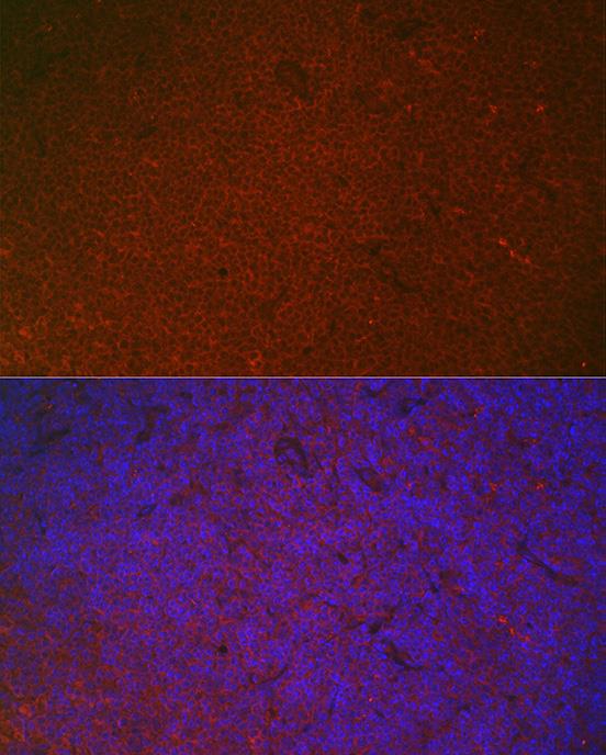
Immunofluorescence analysis of mouse spleen using FCGR2A Rabbit pAb at dilution of 1:100 (40x lens). Blue: DAPI for nuclear staining.

No documents available.