
FGF2

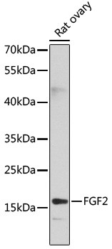
Western blot analysis of extracts of rat ovary, using FGF2 antibody at 1:1000 dilution.
Secondary antibody: HRP Goat Anti-Rabbit IgG (H+L) at 1:10000 dilution.
Lysates/proteins: 25ug per lane.
Blocking buffer: 3% nonfat dry milk in TBST.
Detection: ECL West Pico Kit 
Exposure time: 30s.

Western blot analysis of extracts of rat ovary, using FGF2 antibody at 1:1000 dilution. Secondary antibody: HRP Goat Anti-Rabbit IgG (H+L) at 1:10000 dilution. Lysates/proteins: 25ug per lane. Blocking buffer: 3% nonfat dry milk in TBST. Detection: ECL West Pico Kit Exposure time: 30s.

Catalog Number:AB-90101
Conjugate:Unconjugated
Size:100 ug
Concentration:1mg/ml
Host:Rb
Isotype:IgG
Clone:POLY
Immunogen:Recombinant fusion protein containing a sequence corresponding to amino acids 143-288 of human FGF2
Reactivity:Hu,Ms,Rt
Applications:Western Blot : 1:500-1:2,000 Immunofluorescence/Immunocytochemistry: 1:50- 1:200 Immunohistochemsitry (paraffin, formalin, frozen Tissues): 1:50-1:200
Molecular Weight:20kDa
Purification:Aff. Pur.
Form:Liquid

Applications

Western Blot : 1:500-1:2,000 Immunofluorescence/Immunocytochemistry: 1:50- 1:200 Immunohistochemsitry (paraffin, formalin, frozen Tissues): 1:50-1:200

Immunogen

Recombinant fusion protein containing a sequence corresponding to amino acids 143-288 of human FGF2

Target Background

The protein encoded by this gene is a member of the fibroblast growth factor (FGF) family. FGF family members bind heparin and possess broad mitogenic and angiogenic activities. This protein has been implicated in diverse biological processes, such as limb and nervous system development, wound healing, and tumor growth. The mRNA for this gene contains multiple polyadenylation sites, and is alternatively translated from non-AUG (CUG) and AUG initiation codons, resulting in five different isoforms with distinct properties. The CUG-initiated isoforms are localized in the nucleus and are responsible for the intracrine effect, whereas, the AUG-initiated form is mostly cytosolic and is responsible for the paracrine and autocrine effects of this FGF.

Storage

Storage for short term at 4°C recommended, for longer term at -20°C, minimize freeze/thaw cycles

Buffer

Liquid in PBS with 0,02 % Sodium Azide and 50% glycerol pH 7.3.

Western blot analysis of extracts of rat ovary, using FGF2 antibody at 1:1000 dilution. Secondary antibody: HRP Goat Anti-Rabbit IgG (H+L) at 1:10000 dilution. Lysates/proteins: 25ug per lane. Blocking buffer: 3% nonfat dry milk in TBST. Detection: ECL West Pico Kit Exposure time: 30s.

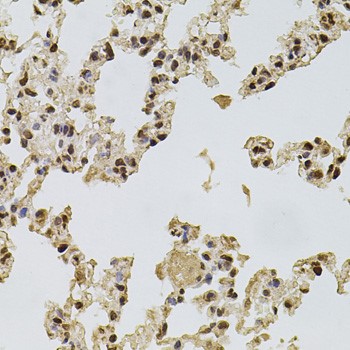
Immunohistochemistry of paraffin-embedded rat lung using FGF2 antibody at dilution of 1:100 (40x lens)

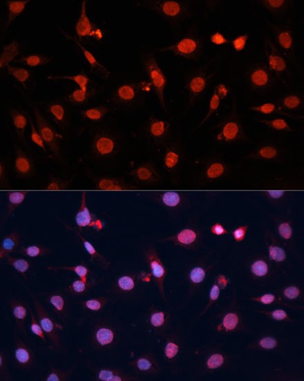
Immunofluorescence analysis of C6 cells using FGF2 Polyclonal Antibody at dilution of 1:100 (40x lens). Blue: DAPI for nuclear staining.

No documents available.