FGFR1 Rabbit Polyclonal Antibody

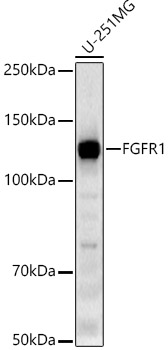
Western blot analysis of U-251MG cells, using FGFR1 antibody at 1:10000 dilution.
Secondary antibody: HRP Goat Anti-Rabbit IgG (H+L) at 1:10000 dilution.
Lysates/proteins: 25μg per lane.
Blocking buffer: 3% nonfat dry milk in TBST.
Detection: ECL West Pico Plus.  
Exposure time: 180s.

Western blot analysis of U-251MG cells, using FGFR1 antibody at 1:10000 dilution. Secondary antibody: HRP Goat Anti-Rabbit IgG (H+L) at 1:10000 dilution. Lysates/proteins: 25μg per lane. Blocking buffer: 3% nonfat dry milk in TBST. Detection: ECL West Pico Plus. Exposure time: 180s.

Catalog Number:AB-84494
Conjugate:Unconjugated
Size:100 ug
Concentration:1mg/ml
Host:Rabbit
Isotype:IgG
Clone:POLY
Immunogen:A synthetic peptide corresponding to a sequence within amino acids 600-700 of human FGFR1
Reactivity:Human, Mouse, Rat
Applications:Westerm Blot: 1:2000 - 1:10000 Immunohistochemistry(paraffin-embedded tissues): 1:50 - 1:200 Immunofluorescnce: 1:50 - 1:200 Immunocytochemistry:1:50 - 1:200 Flow Cytometry: 1:25
Molecular Weight:122 kDa
Purification:Affinity purification
Form:Liquid
Synonyms:CEK; FLG; HH2; OGD; ECCL; FLT2; KAL2; BFGFR; CD331; FGFBR; FLT-2; HBGFR; N-SAM; FGFR-1; HRTFDS; bFGF-R-1

Applications

Westerm Blot: 1:2000 - 1:10000 Immunohistochemistry(paraffin-embedded tissues): 1:50 - 1:200 Immunofluorescnce: 1:50 - 1:200 Immunocytochemistry:1:50 - 1:200 Flow Cytometry: 1:25

Immunogen

A synthetic peptide corresponding to a sequence within amino acids 600-700 of human FGFR1
Target Background

The protein encoded by this gene is a member of the fibroblast growth factor receptor (FGFR) family, where amino acid sequence is highly conserved between members and throughout evolution. FGFR family members differ from one another in their ligand affinities and tissue distribution. A full-length representative protein consists of an extracellular region, composed of three immunoglobulin-like domains, a single hydrophobic membrane-spanning segment and a cytoplasmic tyrosine kinase domain. The extracellular portion of the protein interacts with fibroblast growth factors, setting in motion a cascade of downstream signals, ultimately influencing mitogenesis and differentiation. This particular family member binds both acidic and basic fibroblast growth factors and is involved in limb induction. Mutations in this gene have been associated with Pfeiffer syndrome, Jackson-Weiss syndrome, Antley-Bixler syndrome, osteoglophonic dysplasia, and autosomal dominant Kallmann syndrome 2. Chromosomal aberrations involving this gene are associated with stem cell myeloproliferative disorder and stem cell leukemia lymphoma syndrome. Alternatively spliced variants which encode different protein isoforms have been described; however, not all variants have been fully characterized.

Synonyms:CEK; FLG; HH2; OGD; ECCL; FLT2; KAL2; BFGFR; CD331; FGFBR; FLT-2; HBGFR; N-SAM; FGFR-1; HRTFDS; bFGF-R-1

Storage

Store at -20℃. Avoid freeze / thaw cycles.

Buffer

PBS with 0.05% proclin300,0.05% BSA,50% glycerol,pH7.3.

Western blot analysis of U-251MG cells, using FGFR1 antibody at 1:10000 dilution. Secondary antibody: HRP Goat Anti-Rabbit IgG (H+L) at 1:10000 dilution. Lysates/proteins: 25μg per lane. Blocking buffer: 3% nonfat dry milk in TBST. Detection: ECL West Pico Plus. Exposure time: 180s.

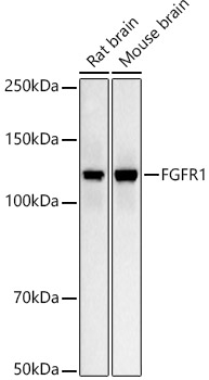
Western blot analysis of various lysates, using FGFR1 antibody at 1:10000 dilution. Secondary antibody: HRP Goat Anti-Rabbit IgG (H+L) at 1:10000 dilution. Lysates/proteins: 25μg per lane. Blocking buffer: 3% nonfat dry milk in TBST. Detection: ECL West Pico Plus. Exposure time: 10s.

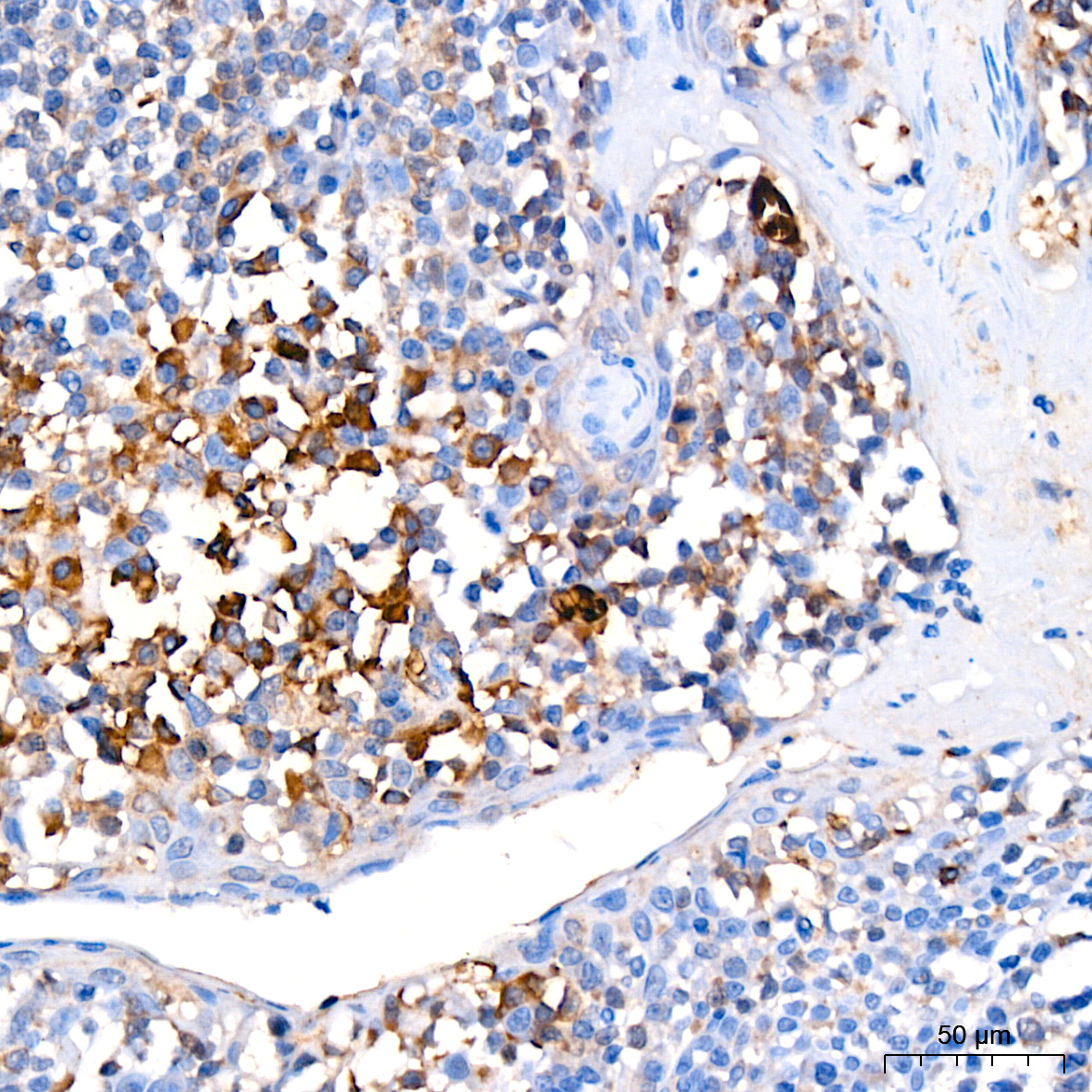
Immunohistochemistry analysis of paraffin-embedded human tonsil using FGFR1 Rabbit pAb at dilution of 1:200 (40x lens).Perform high pressure antigen retrieval with 10 mM citrate buffer pH 6.0 before commencing with IHC staining protocol.

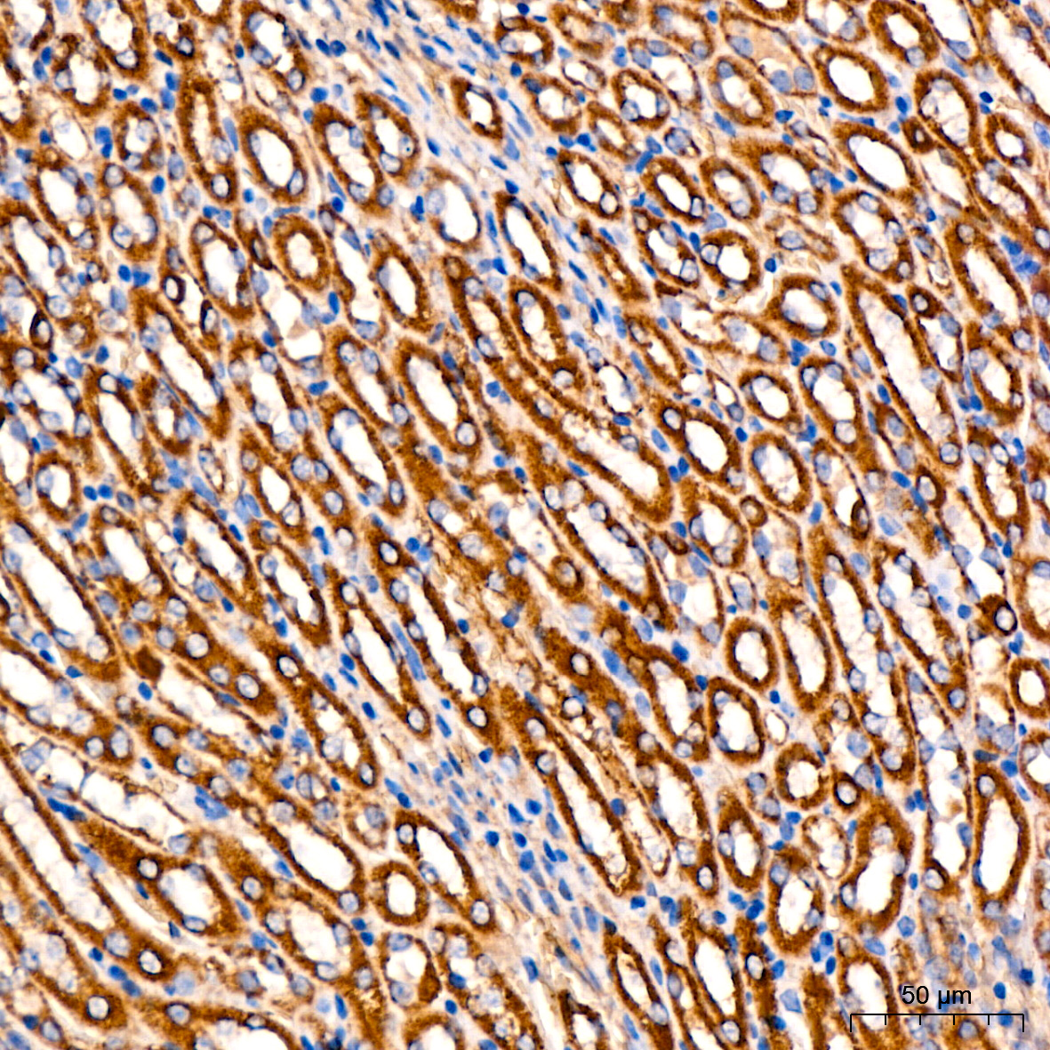
Immunohistochemistry analysis of paraffin-embedded mouse kidney using FGFR1 Rabbit pAb at dilution of 1:200 (40x lens).Perform high pressure antigen retrieval with 10 mM citrate buffer pH 6.0 before commencing with IHC staining protocol.

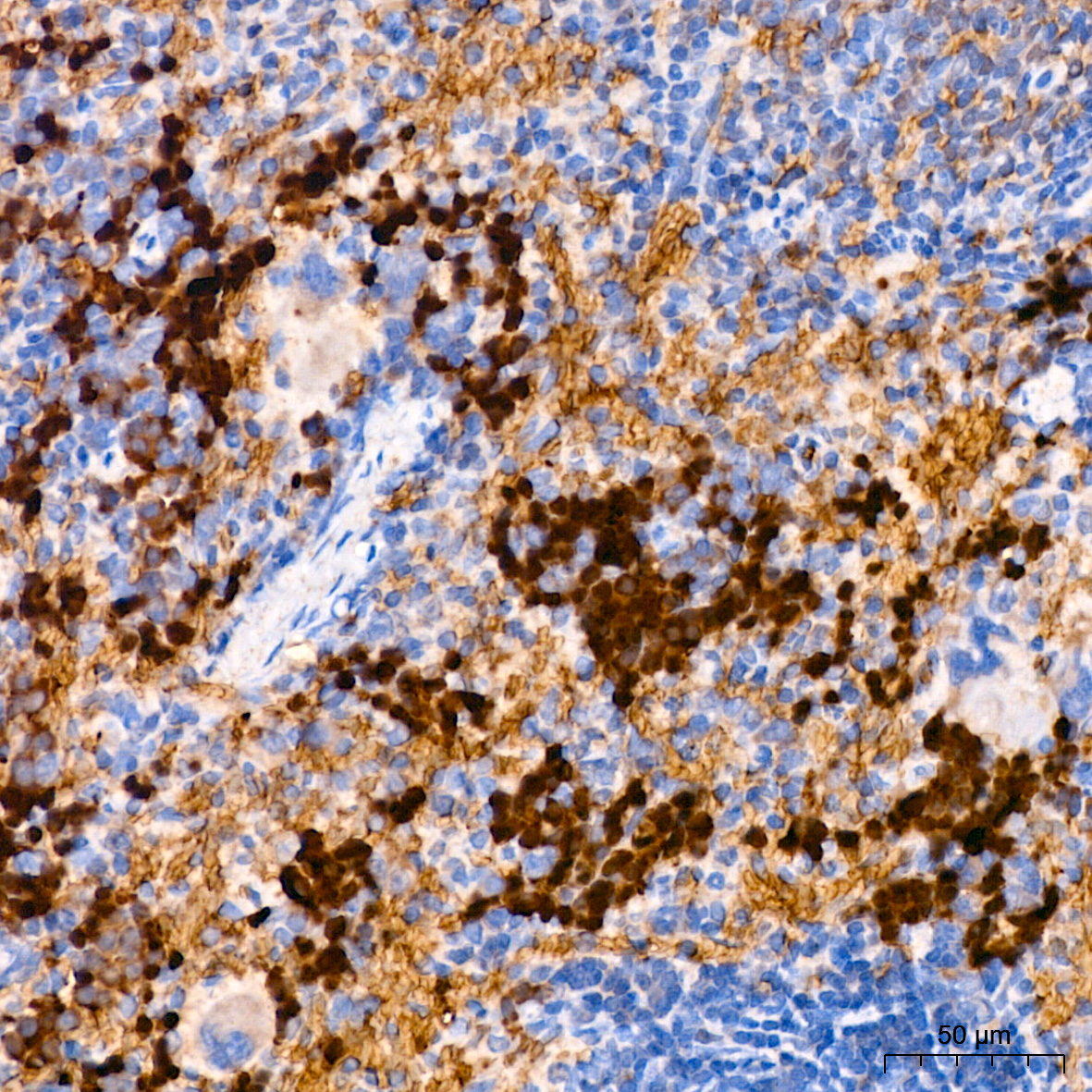
Immunohistochemistry analysis of paraffin-embedded rat spleen using FGFR1 Rabbit pAb at dilution of 1:200 (40x lens).Perform high pressure antigen retrieval with 10 mM citrate buffer pH 6.0 before commencing with IHC staining protocol.

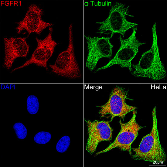
Confocal imaging of HeLa cells using FGFR1 Rabbit pAb dilution 1:200)(Red). The cells were counterstained with α-Tubulin Mouse mAb (AC012,dilution 1:400) (Green). DAPI was used for nuclear staining (blue). Objective: 100x.

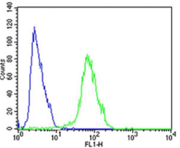
Flow cytometric analysis of SH-SY5Y cells using FGFR1 Antibody (Center)(green) compared to an isotype control of rabbit IgG(blue). FGFR1 was diluted at 1:25 dilution. An Alexa Fluor® 488 goat anti-rabbit lgG at 1:400 dilution was used as the secondary antibody.

No documents available.