FGFR4 Rabbit Monoclonal Antibody

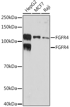
Western blot analysis of extracts of various cell lines, using FGFR4 Rabbit mAb at 1:1000
dilution.
Secondary antibody: HRP Goat Anti-Rabbit IgG (H+L) at 1:10000 dilution.
Lysates/proteins: 25ug per lane.
Blocking buffer: 3% nonfat dry milk in TBST.
Detection: ECL West Pico Plus. 
Exposure time: 3min.

Western blot analysis of extracts of various cell lines, using FGFR4 Rabbit mAb at 1:1000 dilution. Secondary antibody: HRP Goat Anti-Rabbit IgG (H+L) at 1:10000 dilution. Lysates/proteins: 25ug per lane. Blocking buffer: 3% nonfat dry milk in TBST. Detection: ECL West Pico Plus. Exposure time: 3min.

Catalog Number:MAB-94770
Conjugate:Unconjugated
Size:100 ug
Concentration:1mg/ml
Host:Rabbit
Isotype:IgG
Clone:ARC1474
Immunogen:A synthetic peptide corresponding to a sequence within amino acids 700-802 of human FGFR4.
Reactivity:Human, Mouse
Applications:Western Blot: 1:500 - 1:2000 Immunohistochemistry (Paraffin-embedded tissues): 1:50 - 1:200
Molecular Weight:95KDa/125KDa
Purification:Affinity purification
Form:Liquid
Synonyms:TKF; JTK2; CD334

Applications

Western Blot: 1:500 - 1:2000 Immunohistochemistry (Paraffin-embedded tissues): 1:50 - 1:200

Immunogen

A synthetic peptide corresponding to a sequence within amino acids 700-802 of human FGFR4.
Target Background

The protein encoded by this gene is a tyrosine kinase and cell surface receptor for fibroblast growth factors. The encoded protein is involved in the regulation of several pathways, including cell proliferation, cell differentiation, cell migration, lipid metabolism, bile acid biosynthesis, vitamin D metabolism, glucose uptake, and phosphate homeostasis. This protein consists of an extracellular region, composed of three immunoglobulin-like domains, a single hydrophobic membrane-spanning segment, and a cytoplasmic tyrosine kinase domain. The extracellular portion interacts with fibroblast growth factors, setting in motion a cascade of downstream signals, ultimately influencing mitogenesis and differentiation.

Synonyms:TKF; JTK2; CD334

Storage

Store at -20℃. Avoid freeze / thaw cycles.

Buffer

PBS with 0.02% sodium azide,0.05% BSA,50% glycerol,pH7.3.

Western blot analysis of extracts of various cell lines, using FGFR4 Rabbit mAb at 1:1000 dilution. Secondary antibody: HRP Goat Anti-Rabbit IgG (H+L) at 1:10000 dilution. Lysates/proteins: 25ug per lane. Blocking buffer: 3% nonfat dry milk in TBST. Detection: ECL West Pico Plus. Exposure time: 3min.

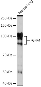
Western blot analysis of extracts of Mouse lung, using FGFR4 Rabbit mAb at 1:1000 dilution. Secondary antibody: HRP Goat Anti-Rabbit IgG (H+L) at 1:10000 dilution. Lysates/proteins: 25ug per lane. Blocking buffer: 3% nonfat dry milk in TBST. Detection: ECL West Pico Plus. Exposure time: 3min.

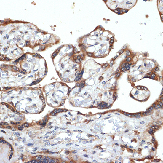
Immunohistochemistry of paraffinembedded human placenta using FGFR4 Rabbit mAb at dilution of 1:100 (40x lens).Perform microwave antigen retrieval with 10 mM Tris/EDTA buffer pH 9.0 before commencing with IHC staining protocol.

No documents available.