Filamin A Rabbit Monoclonal Antibody

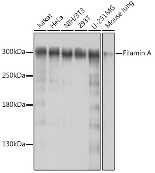
Western blot analysis of extracts of various cell lines, using Filamin A antibody at 1:1000
dilution.
Secondary antibody: HRP Goat Anti-Rabbit IgG (H+L) at 1:10000 dilution.
Lysates/proteins: 25μg per lane.
Blocking buffer: 3% nonfat dry milk in TBST.
Detection: ECL West Pico Plus. 
Exposure time: 30s.

Western blot analysis of extracts of various cell lines, using Filamin A antibody at 1:1000 dilution. Secondary antibody: HRP Goat Anti-Rabbit IgG (H+L) at 1:10000 dilution. Lysates/proteins: 25μg per lane. Blocking buffer: 3% nonfat dry milk in TBST. Detection: ECL West Pico Plus. Exposure time: 30s.

Catalog Number:MAB-94782
Conjugate:Unconjugated
Size:100 ug
Concentration:1mg/ml
Host:Rabbit
Isotype:IgG
Clone:PMS/317
Immunogen:Recombinant fusion protein containing a sequence corresponding to amino acids 2348-2647 of human Filamin A
Reactivity:Human, Mouse, Rat
Applications:Western Blot: 1:500 - 1:2000 Immunohistochemistry (paraffin-embedded tissues): 1:50 - 1:200 Immunofluorescence: 1:50 - 1:200 Immunocytcohemistry: 1:50 - 1:200
Molecular Weight:300kDa
Purification:Affinity purification
Form:Liquid
Synonyms:FLN; FMD; MNS; OPD; ABPX; CSBS; CVD1; FGS2; FLN1; NHBP; OPD1; OPD2; XLVD; XMVD; FLN-A; ABP-280

Applications

Western Blot: 1:500 - 1:2000 Immunohistochemistry (paraffin-embedded tissues): 1:50 - 1:200 Immunofluorescence: 1:50 - 1:200 Immunocytcohemistry: 1:50 - 1:200

Immunogen

Recombinant fusion protein containing a sequence corresponding to amino acids 2348-2647 of human Filamin A

Target Background

The protein encoded by this gene is an actin-binding protein that crosslinks actin filaments and links actin filaments to membrane glycoproteins. The encoded protein is involved in remodeling the cytoskeleton to effect changes in cell shape and migration. This protein interacts with integrins, transmembrane receptor complexes, and second messengers. Defects in this gene are a cause of several syndromes, including periventricular nodular heterotopias (PVNH1, PVNH4), otopalatodigital syndromes (OPD1, OPD2), frontometaphyseal dysplasia (FMD), Melnick-Needles syndrome (MNS), and X-linked congenital idiopathic intestinal pseudoobstruction (CIIPX). Two transcript variants encoding different isoforms have been found for this gene.

Synonyms:FLN; FMD; MNS; OPD; ABPX; CSBS; CVD1; FGS2; FLN1; NHBP; OPD1; OPD2; XLVD; XMVD; FLN-A; ABP-280

Storage

Store at -20℃. Avoid freeze / thaw cycles.

Buffer

PBS with 0.02% sodium azide,50% glycerol,pH7.3.

Western blot analysis of extracts of various cell lines, using Filamin A antibody at 1:1000 dilution. Secondary antibody: HRP Goat Anti-Rabbit IgG (H+L) at 1:10000 dilution. Lysates/proteins: 25μg per lane. Blocking buffer: 3% nonfat dry milk in TBST. Detection: ECL West Pico Plus. Exposure time: 30s.

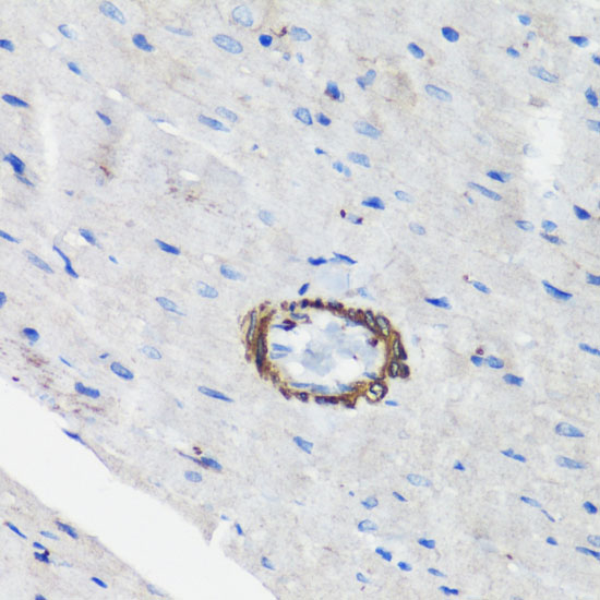
Immunohistochemistry analysis of paraffin-embedded rat heart using Filamin A antibody at dilution of 1:100 (40x lens).Perform microwave antigen retrieval with 10 mM PBS buffer pH 7.2 before commencing with IHC staining protocol.

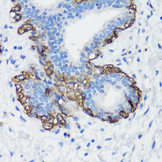
Immunohistochemistry analysis of paraffin-embedded human breast cancer using Filamin A antibody at dilution of 1:100 (40x lens).Perform microwave antigen retrieval with 10 mM PBS buffer pH 7.2 before commencing with IHC staining protocol.

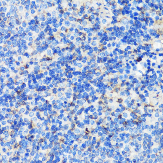
Immunohistochemistry analysis of paraffin-embedded mouse spleen using Filamin A antibody at dilution of 1:100 (40x lens).Perform microwave antigen retrieval with 10 mM PBS buffer pH 7.2 before commencing with IHC staining protocol.

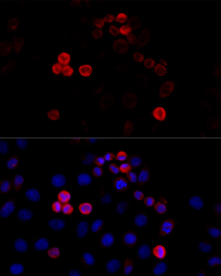
Immunofluorescence analysis of HeLa cells using Filamin A antibody at dilution of 1:100 (40x lens). B lue: DAPI for nuclear staining.

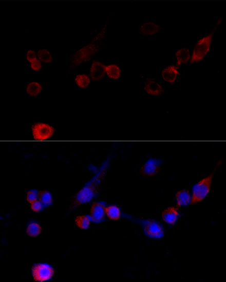
Immunofluorescence analysis of NIH/3T3 cells using Filamin A antibody at dilution of 1:100 (40x lens). Blue: DAPI for nuclear staining.

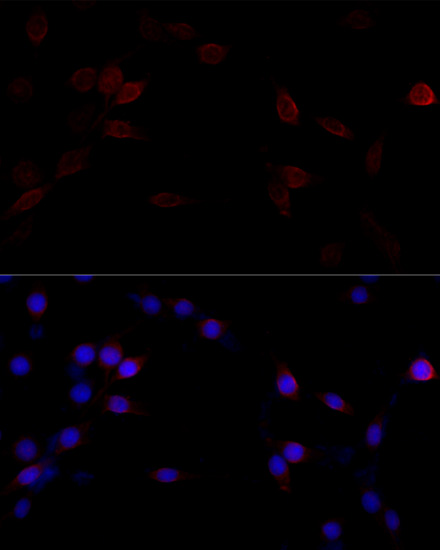
Immunofluorescence analysis of PC-12 cells using Filamin A antibody at dilution of 1:100 (40x lens). Blue: DAPI for nuclear staining.

No documents available.