
FITC Anti-Human CD1a Antibody[OKT-6]
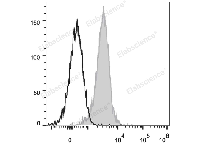
Human T lymphoblastic leukemia cell line Molt-4 are stained with FITC Anti-Human CD1a Antibody (filled gray histogram) or Mouse IgG1 Isotype Control FITC (empty black histogram).
| Catalog Number: | MAB1126C |
| Conjugate: | FITC is designed to be excited by the Blue laser (488 nm) and detected using an optical filter centered near 530 nm (e.g., a 525/40 nm bandpass filter). |
| Size: | 100Tests |
| Concentration: | 5 μL/Test |
| Host: | Mouse |
| Isotype: | Mouse IgG1, κ |
| Clone: | OKT-6 |
| Reactivity: | Human |
| Applications: | FCM - Each lot of this antibody is quality control tested by flow cytometric analysis. <b style="color:#0070ca">The amount of the reagent is suggested to be used 5 µL of antibody per test (million cells in 100 µL staining volume or per 100 µL of whole blood).</b> Please check your vial before the experiment. Since applications vary, the appropriate dilutions must be determined for individual use. |
| Form: | Liquid |
CD1a is a 49 kD member of the immunoglobulin superfamily also known as T6 and R4. It is a type I membrane glycoprotein with structural similarities to MHC class I and is non-covalently associated with β2-microglobulin. CD1a plays a role in non-peptide glycolipid antigen presentation to CD1-restricted T cells. It is expressed on cortical double positive and single positive thymocytes, Langerhans cells, and dendritic cells. In addition to antigen presentation, CD1a has been implicated in thymic T cell development.
Applications
FCM - Each lot of this antibody is quality control tested by flow cytometric analysis. <b style="color:#0070ca">The amount of the reagent is suggested to be used 5 µL of antibody per test (million cells in 100 µL staining volume or per 100 µL of whole blood).</b> Please check your vial before the experiment. Since applications vary, the appropriate dilutions must be determined for individual use.