FOXP3 Rabbit Monoclonal Antibody

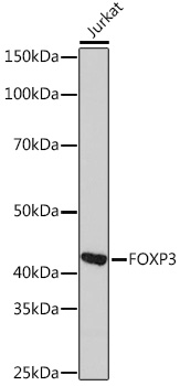
Western blot analysis of extracts of Jurkat cells, using FOXP3 Rabbit mAb  at 1:1000 dilution.
Secondary antibody: HRP Goat Anti-Rabbit IgG (H+L) at 1:10000 dilution.
Lysates/proteins: 25ug per lane.
Blocking buffer: 3% nonfat dry milk in TBST.
Detection: ECL West Pico Plus.
Exposure time: 10s.

Western blot analysis of extracts of Jurkat cells, using FOXP3 Rabbit mAb  at 1:1000 dilution. Secondary antibody: HRP Goat Anti-Rabbit IgG (H+L) at 1:10000 dilution. Lysates/proteins: 25ug per lane. Blocking buffer: 3% nonfat dry milk in TBST. Detection: ECL West Pico Plus. Exposure time: 10s.

Catalog Number:MAB-94759
Conjugate:Unconjugated
Size:100 ug
Concentration:1mg/ml
Host:Rabbit
Isotype:IgG
Clone:ARC1159
Immunogen:Recombinant fusion protein containing a sequence corresponding to amino acids 286-431 of human FOXP3
Reactivity:Human
Applications:Western Blot:                   1:500 - 1:2000 Immunoprecipitation:   1:500 - 1:1000 Immunofluorescence:  1:200-1:500
Molecular Weight:45KDa
Purification:Affinity purification
Form:Liquid
Synonyms:AIID; DIETER; IPEX; JM2; PIDX; XPID

Applications

Western Blot:                   1:500 - 1:2000 Immunoprecipitation:   1:500 - 1:1000 Immunofluorescence:  1:200-1:500

Immunogen

Recombinant fusion protein containing a sequence corresponding to amino acids 286-431 of human FOXP3

Target Background

The protein encoded by this gene is a member of the forkhead/winged-helix family of transcriptional regulators. Defects in this gene are the cause of immunodeficiency polyendocrinopathy, enteropathy, X-linked syndrome (IPEX), also known as X-linked autoimmunity-immunodeficiency syndrome. Alternatively spliced transcript variants encoding different isoforms have been identified.

Synonyms:AIID; DIETER; IPEX; JM2; PIDX; XPID

Storage

Store at -20℃. Avoid freeze / thaw cycles.

Buffer

PBS with 0.02% sodium azide,0.05% BSA,50% glycerol,pH7.3.

Western blot analysis of extracts of Jurkat cells, using FOXP3 Rabbit mAb  at 1:1000 dilution. Secondary antibody: HRP Goat Anti-Rabbit IgG (H+L) at 1:10000 dilution. Lysates/proteins: 25ug per lane. Blocking buffer: 3% nonfat dry milk in TBST. Detection: ECL West Pico Plus. Exposure time: 10s.

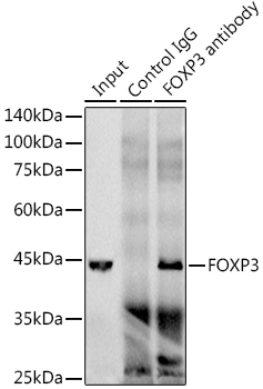
Immunoprecipitation analysis of 300ug extracts of Jurkat cells using 3ug FOXP3 antibody. Western blot was performed from the immunoprecipitate using FOXP3 antibody at a dilution of 1:1000.

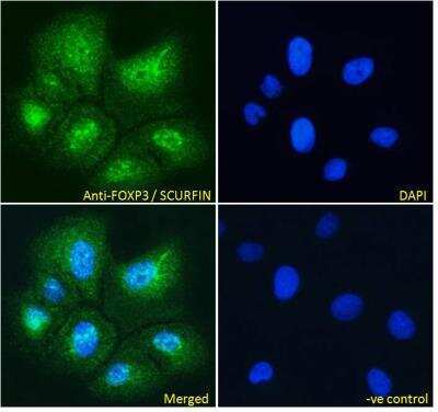
Immunofluorescence: FoxP3 Antibody Paraformaldehyde fixed U2OS cells, permeabilized with 0.15% Triton. Primary incubation 1hr (10 ug/mL) followed by Alexa Fluor 488 secondary antibody (4 ug/mL), showing strong localization to nucleoplasm. The nuclear stain is DAPI (blue). Negative control: Unimmunized goat IgG (10 ug/mL) followed by Alexa Fluor 488 secondary antibody (4 ug/mL).

No documents available.