FOXp3 Rabbit Monoclonal Antibody

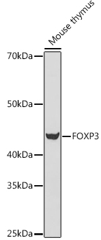
Western blot analysis of extracts of Mouse thymus cells, using FOXP3 antibody  at 1:500 dilution.
Secondary antibody: HRP Goat Anti-Rabbit IgG (H+L) at 1:10000 dilution.
Lysates/proteins: 25ug per lane.
Blocking buffer: 3% nonfat dry milk in TBST.
Detection: ECL West Pico Plus.
Exposure time: 180s.

Western blot analysis of extracts of Mouse thymus cells, using FOXP3 antibody  at 1:500 dilution. Secondary antibody: HRP Goat Anti-Rabbit IgG (H+L) at 1:10000 dilution. Lysates/proteins: 25ug per lane. Blocking buffer: 3% nonfat dry milk in TBST. Detection: ECL West Pico Plus. Exposure time: 180s.

Catalog Number:MAB-94761
Conjugate:Unconjugated
Size:100 ug
Concentration:1mg/ml
Host:Rabbit
Isotype:IgG
Clone:ARC505
Immunogen:Recombinant fusion protein containing a sequence corresponding to amino acids 206-300 of human FOXP3
Reactivity:Human, Mouse, Rat
Applications:Western Blot:        1:100 - 1:500 Immunohistochemistry: 1:50-1:200
Molecular Weight:47KDa
Purification:Affinity purification
Form:Liquid
Synonyms:FOXP3;AIID;DIETER;IPEX;JM2;PIDX;XPID

Applications

Western Blot:        1:100 - 1:500 Immunohistochemistry: 1:50-1:200

Immunogen

Recombinant fusion protein containing a sequence corresponding to amino acids 206-300 of human FOXP3

Target Background

The protein encoded by this gene is a member of the forkhead/winged-helix family of transcriptional regulators. Defects in this gene are the cause of immunodeficiency polyendocrinopathy, enteropathy, X-linked syndrome (IPEX), also known as X-linked autoimmunity-immunodeficiency syndrome. Alternatively spliced transcript variants encoding different isoforms have been identified.

Synonyms:FOXP3;AIID;DIETER;IPEX;JM2;PIDX;XPID

Storage

Store at -20℃. Avoid freeze / thaw cycles.

Buffer

PBS with 0.01% thiomersal,50% glycerol,pH7.3.

Western blot analysis of extracts of Mouse thymus cells, using FOXP3 antibody  at 1:500 dilution. Secondary antibody: HRP Goat Anti-Rabbit IgG (H+L) at 1:10000 dilution. Lysates/proteins: 25ug per lane. Blocking buffer: 3% nonfat dry milk in TBST. Detection: ECL West Pico Plus. Exposure time: 180s.

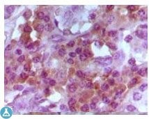
Immunohistochemistry (IHC) analysis of paraffin-embedded Human Lymphocyto tissue, showing cytoplasmic and nuclear localization with DAB staining using FoxP3 Monoclonal Antibody.

No documents available.