GAPDH Mouse Monoclonal Antibody

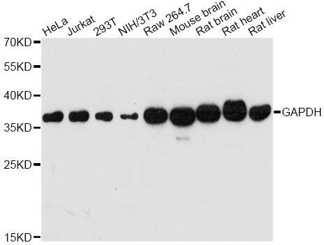
Western blot analysis of extracts of various cell lines, using GAPDH antibody at 1:10,000 dilution.
Secondary antibody: HRP Goat Anti-Mouse IgG (H+L) at 1:10,000 dilution.
Lysates/proteins: 25ug per lane.
Blocking buffer: 3% nonfat dry milk in TBST.
Detection: ECL Basic Kit 
Exposure time:60s

Western blot analysis of extracts of various cell lines, using GAPDH antibody at 1:10,000 dilution. Secondary antibody: HRP Goat Anti-Mouse IgG (H+L) at 1:10,000 dilution. Lysates/proteins: 25ug per lane. Blocking buffer: 3% nonfat dry milk in TBST. Detection: ECL Basic Kit Exposure time:60s

Catalog Number:MAB-10578
Conjugate:Unconjugated
Size:100 ug
Concentration:1mg/ml
Host:Ms
Isotype:IgG1
Clone:6C5
Immunogen:Recombinant protein of human GAPDH
Reactivity:Hu, Ms, Rt
Applications:Western Blot: 1:5,000-1:50,000 Immunofluorescence 1:200 Immunocytochemistry: 1:50-200 Immunohistochemistry (paraffin embedded tissues):1:50-200
Molecular Weight:36kDa
Purification:Aff. Pur.
Form:Liquid

Applications

Western Blot: 1:5,000-1:50,000 Immunofluorescence 1:200 Immunocytochemistry: 1:50-200 Immunohistochemistry (paraffin embedded tissues):1:50-200

Immunogen

Recombinant protein of human GAPDH
Target Background

Glyceraldehyde-3-phosphate dehydrogenase (GAPDH) catalyzes the phosphorylation of glyceraldehyde-3-phosphate during glycolysis. GAPDH participates in nuclear events including transcription, binding RNA, RNA transportation, DNA replication, DNA repair and apoptosis. Being stably and constitutively expressed at high levels in most tissues and cells, GAPDH is considered a housekeeping protein. It was widely used as a control for RT-PCR and also loading control in electrophoresis and Western blotting. GAPDH is normally expressed in cellular cytoplasm or membrane, but can occasionally translocated to the nucleus post modification such as S-nitrosylation. This antibody is a mouse monoclonal antibody raised against full length GAPDH of human origin. It can recognize the 36kda GAPDH protein in most cells/tissues. Please note that some physiological factors, such as hypoxia and diabetes, increase GAPDH expression in certain cell types.

Storage

Store at -20℃. Avoid freeze / thaw cycles.

Buffer

PBS with 0.02% sodium azide, 50% glycerol, pH7.3.

Western blot analysis of extracts of various cell lines, using GAPDH antibody at 1:10,000 dilution. Secondary antibody: HRP Goat Anti-Mouse IgG (H+L) at 1:10,000 dilution. Lysates/proteins: 25ug per lane. Blocking buffer: 3% nonfat dry milk in TBST. Detection: ECL Basic Kit Exposure time:60s

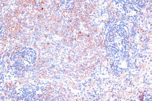
Immunohistochemistry of paraffin-embedded rat spleen using GAPDH antibody at dilution of 1:100

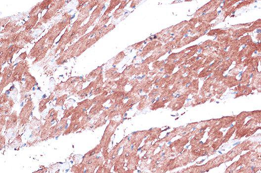
Immunohistochemistry of paraffin-embedded rat heart using GAPDH 1:100

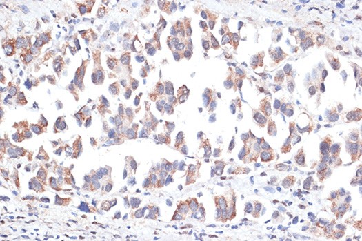
Immunohistochemistry of paraffin-embedded human lung cancer using GAPDH antibody of 1:100

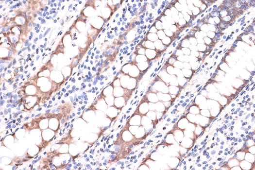
Immunohistochemistry of paraffin-embedded human colon using GAPDH antibody at dilution of 1:100

Immunohistochemistry of paraffin-embedded mouse spinal cord using GAPDH antibody at dilution of 1:100

No documents available.