GFAP

Western blot analysis of whole tissue lysates using mouse mAb to GFAP, dilution 1:1,000, in green:                 [1] protein standard (red), [2] rat brain, [3] rat spinal cord, [4] mouse brain, [5] mouse spinal cord.                        The strong band at about 50kDa corresponds to the GFAP protein

Western blot analysis of whole tissue lysates using mouse mAb to GFAP, dilution 1:1,000, in green: [1] protein standard (red), [2] rat brain, [3] rat spinal cord, [4] mouse brain, [5] mouse spinal cord. The strong band at about 50kDa corresponds to the GFAP protein

Catalog Number:MAB-94373
Conjugate:Unconjugated
Size:100 ug
Concentration:1mg/ml
Host:Ms
Isotype:IgG
Clone:G-A-5
Immunogen:Purified porcine spinal cord GFAP
Reactivity:Hu, Ms, Rt, Cw, Pg, Ho
Applications:Western blot: 1:2,500 Immunohistochemistry (Paraffin embedded tissues):1:500 Immunohistochemistry (Frozen Tissues) 1:500 - 1,000 Immunofluorescence : 1: :500 – 1: 1,000 Immunocytochemistry: 1: 500 – 1: 1,000
Purification:Aff. Pur.
Form:Liquid

Applications

Western blot: 1:2,500 Immunohistochemistry (Paraffin embedded tissues):1:500 Immunohistochemistry (Frozen Tissues) 1:500 - 1,000 Immunofluorescence : 1: :500 – 1: 1,000 Immunocytochemistry: 1: 500 – 1: 1,000

Immunogen

Purified porcine spinal cord GFAP
Target Background

Glial Fibrillary Acidic Protein (GFAP) was discovered by Amico Bignami and coworkers as a major fibrous protein of multiple sclerosis plaques (1). It was subsequently found to be a member of the 10nm or intermediate filament protein family, specifically the intermediate filament protein family Class III, which also includes peripherin, desmin and vimentin. The GFAP protein runs on gels as a ~50kDa protein, usually associated with somewhat lower molecule weight bands which are alternate transcripts from the single gene. The HGNC nomenclature for this protein is, perhaps not surprisingly, GFAP. GFAP is strongly and specifically expressed in astrocytes and certain other astroglia in the central nervous system, in satellite cells in peripheral ganglia, and in non-myelinating Schwann cells in peripheral nerves (2,3). It is also a component of neural stem cells.

Storage

At 4°C short term or -20°C long term. Avoid repeated freezing and thawing.

Buffer

Supplied in PBS 50% glycerol, 5 mM Sodium Azide

Western blot analysis of whole tissue lysates using mouse mAb to GFAP, dilution 1:1,000, in green: [1] protein standard (red), [2] rat brain, [3] rat spinal cord, [4] mouse brain, [5] mouse spinal cord. The strong band at about 50kDa corresponds to the GFAP protein

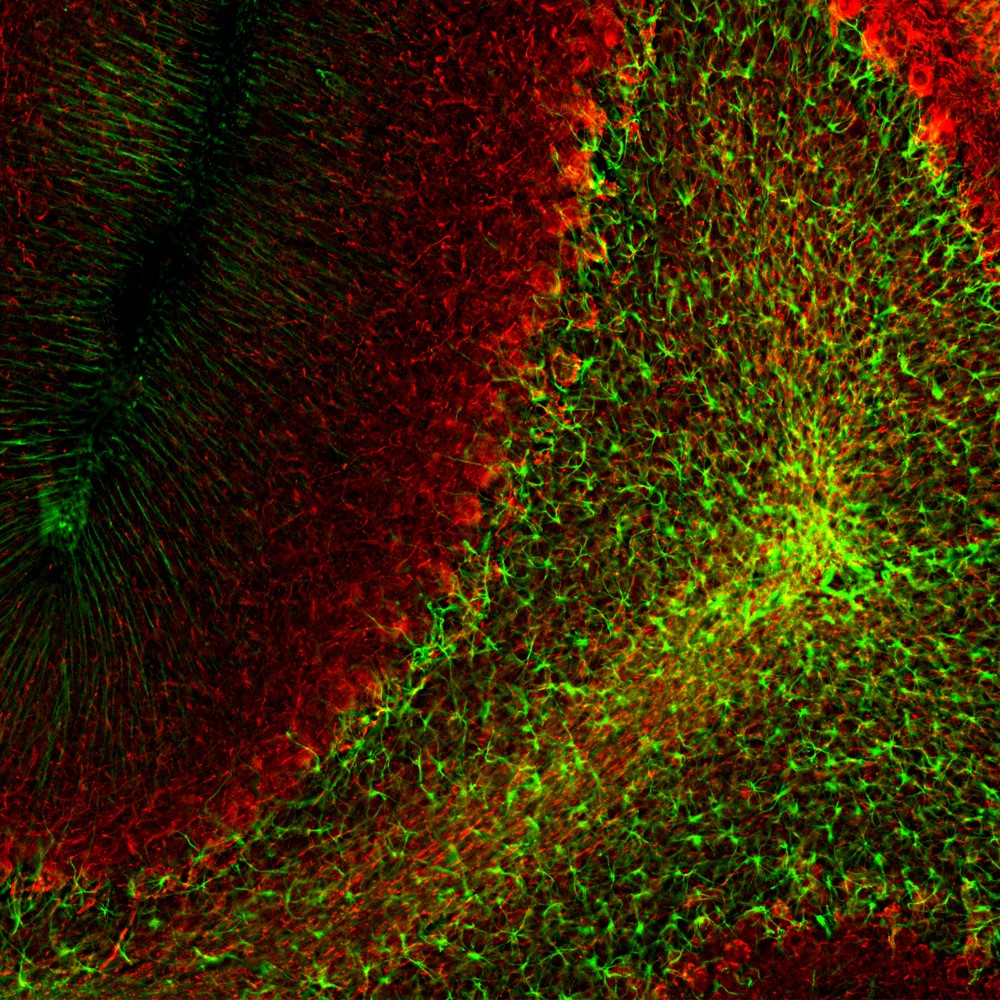
Immunofluorescent analysis of rat cerebellum section stained with mouse mAb to GFAP, dilution 1:1,000, in green, costained with rabbit pAb to neurofilament NF-L dilution 1:1,000, in red. Following transcardial perfusion with 4% paraformaldehyde, brain was post fixed for 24 hours, cut to 45μM, and free-floating sections were stained with antibodies. MAB-94373 stains a network of astroglial cells, while the NF-L antibody labels neuronal cells and their processes

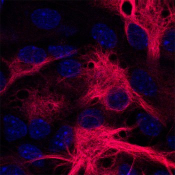
Mouse monoclonal to GFAP [GF5] was used in fixed murine cultures (mixed neurons/glia) at 1/100 overnight at 4°C. A secondary goat anti-mouse antibody was used for detection (Alexa Fluor 568; 1/400). Microscopy revealed diffuse cytosolic labelling. Coounter staining with TO-PRO-3 (Molecular Probes; 660nm (converted here to blue colour) was used to identify the nucleus. The “fibrous” anti-GFAP staining of murine mixed cultures is typical of what is expected.

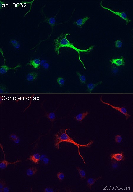
GFAP antibody [GF5] - Astrocyte Marker MAB-94373 immunocytochemical detection in stimulated Cor1 cells. Stimulated Cor1 cells were fixed in formaldehyde, permeabilized, blocked in 1% BSA for 10 mins @ rt°C. Primary Antibody MAB-94373 incubated at 1/1500 for 2 hours in TBS/BSA/azide/0.3% triton. Secondary Antibody: anti mouse IgG Conjugated to: Alexa Fluor® 488 (1/1000).

No documents available.