GFAP Rabbit Polyclonal Antibody

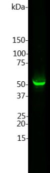
Left:  Western blot of whole rat 
cerebellum homogenate stained  with GFAP, at dilution of 1:2,500. A prominent band running with an apparent SDS-PAGE molecular weight of ~50kDa corresponds to rodent
GFAP. A lower band at ~45kDa is derived from the GFAP molecule.

Left: Western blot of whole rat cerebellum homogenate stained with GFAP, at dilution of 1:2,500. A prominent band running with an apparent SDS-PAGE molecular weight of ~50kDa corresponds to rodent GFAP. A lower band at ~45kDa is derived from the GFAP molecule.

Catalog Number:AB-10682
Conjugate:Unconjugated
Size:100 ul
Concentration:1mg/ml
Host:Rabbit
Isotype:IgG
Clone:POLY
Immunogen:Purified bovine full length protein
Reactivity:Hu, Ms, Rt, Ct, Mamm.
Applications:Western Blot: 1: 2,500 is recommendedImmunocytochemistry: on cell in tissue cultures at 1:1,000 using fluorescent secondary antibody Immunofluorescence: 1:1,000Immunohistochemistry: in tissue sections (frozen and paraffin-embedded tissues) at 1:500-1:1,000 or 1:2,500 using other enzyme linked methodImmunohistochemistry (frozen): 1:500
Molecular Weight:55kDa
Purification:Serum
Form:Liquid

Applications

Western Blot: 1: 2,500 is recommendedImmunocytochemistry: on cell in tissue cultures at 1:1,000 using fluorescent secondary antibody Immunofluorescence: 1:1,000Immunohistochemistry: in tissue sections (frozen and paraffin-embedded tissues) at 1:500-1:1,000 or 1:2,500 using other enzyme linked methodImmunohistochemistry (frozen): 1:500

Immunogen

Purified bovine full length protein
Target Background

Glial Fibrillary Acidic Protein (GFAP) was discovered by Amico Bignami and coworkers as a majorfibrous protein of multiple sclerosis plaques (1). It was subsequently found to be a member of the 10nm or intermediate filament protein family, specifically the intermediate filament protein family Class III, which also includes peripherin, desmin and vimentin. The GFAP protein runs on gels at ~55kDa protein, usually associated with lower molecule weight bands which are thought to be proteolytic fragments and alternate transcripts from the single gene. GFAP is strongly and specifically expressed in astrocytes and certain other astroglia in the central nervous system, in satellite cells in peripheral ganglia, and in non-myelinating Schwann cells in peripheral nerves. In many damage and disease states GFAP expression is heavily upregulated in astrocytes. In addition neural stem cells frequently strongly express GFAP. Antibodies to GFAP are therefore very useful as markers of astrocytic cells and neural stem cells. In addition many types of brain tumor, presumably derived from astrocytic cells, heavily express GFAP. Finally, Alexander's disease was recently shown to be caused by point mutations in protein coding region of the GFAP gene (2). All forms of Alexander disease are characterized by the presence of Rosenthal fibers, which are GFAP containing cytoplasmic inclusions found in astrocytes. Human, horse, cow, pig, chicken, rat, mouse and other mammalians.

Storage

At 4°-8° C for short term. At -20°C For longer term. Avoid repeated freezing and thawing cycles.

Buffer

Antibody is supplied as an aliquot of serum plus 5mM NaN3

Left: Western blot of whole rat cerebellum homogenate stained with GFAP, at dilution of 1:2,500. A prominent band running with an apparent SDS-PAGE molecular weight of ~50kDa corresponds to rodent GFAP. A lower band at ~45kDa is derived from the GFAP molecule.

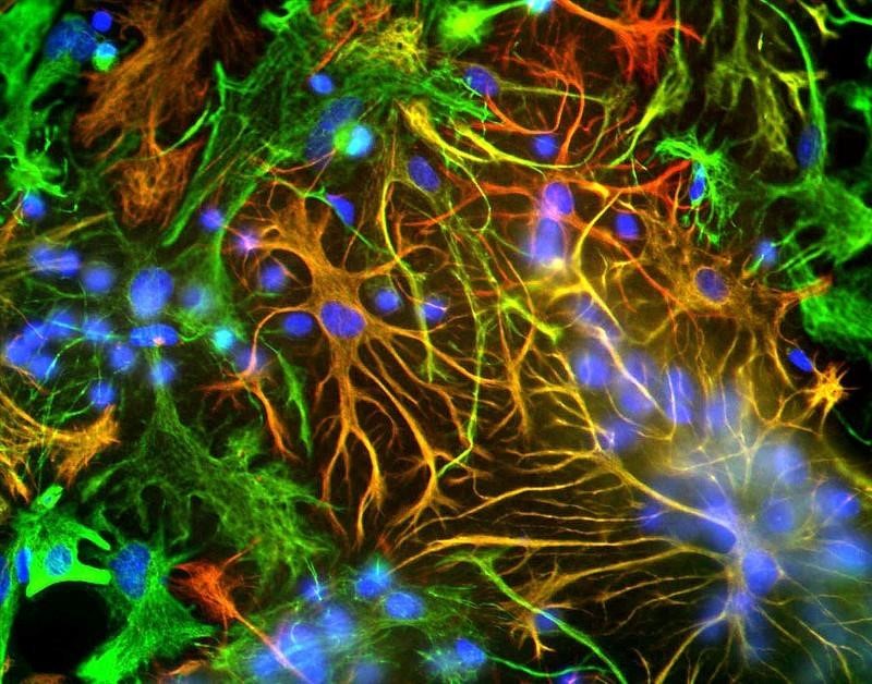
Immunocytochemistry/Immunofluorescence: GFAP Antibody ICC-IF analysis of mixed neuron-glial cultures using GFAP antibody (red) and Vimentin antibody (green). The fibroblastic cells contain only Vimentin and so are green. The astrocytes contain either Vimentin and GFAP (appearing golden) or predominantly GFAP (appearing red). Blue is nuclear DNA stain

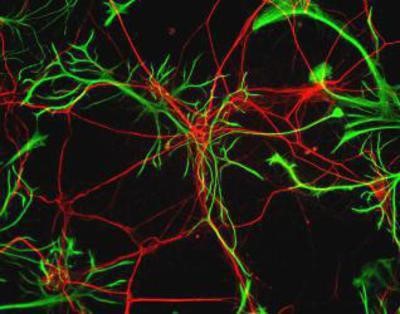
Immunocytochemistry/Immunofluorescence: GFAP Antibody - Rat neurons stained with Neurofilament Heavy antibody (red) and GFAP antibody (green).

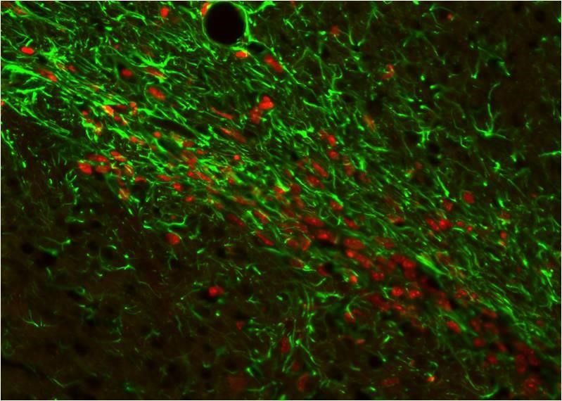
Immunocytochemistry/Immunofluorescence: GFAP Antibody Xenografted mouse brain section : astocyte and human nuclei.

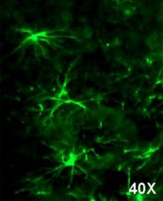
Immunohistochemistry-Frozen: GFAP Antibody Imaging of mouse brain (cortex), 20x magnification

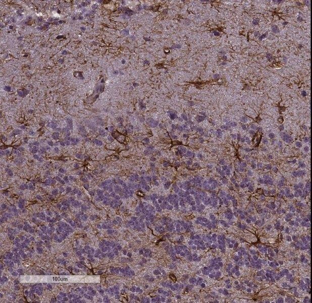
Immunohistochemistry frozen tissues At a 1/500 dilution staining rat spinal cord tissue sections by IHC-Fr. Rats were transcardially perfused with 4% PFA. The tissue was post fixed 1 hour in 4% PFA and then 30% sucrose for three days. 20µm sections were cryostat cut. The primary antibody was incubated with the tissue sections for 18 hours. Bound antibody was detected using an Alexa Fluor 488 conjugated goat anti-rabbit polyclonal

No documents available.