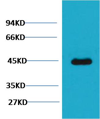
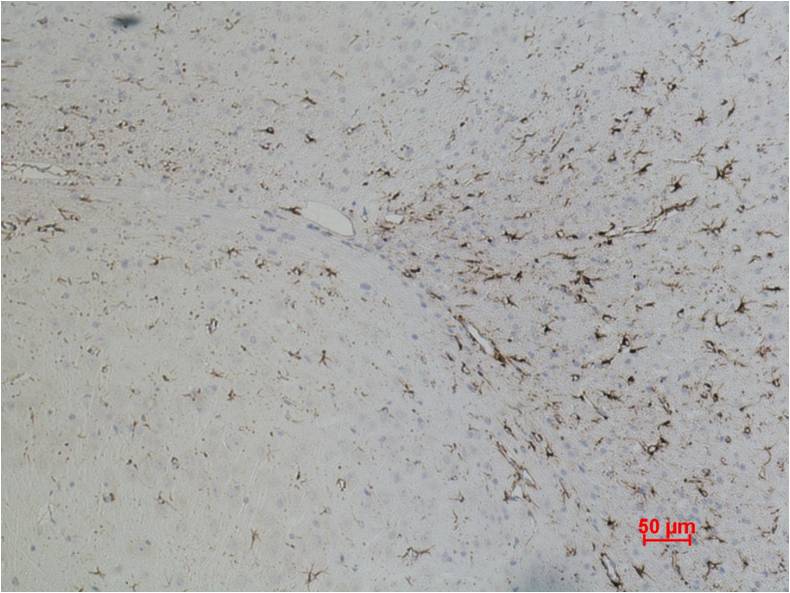
GFAP Mouse mAb
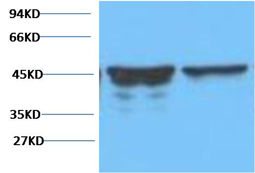
Western blot analysis of Rat Brain Tissue with GFAP mAb diluted at 1:5,000.
| Catalog Number: | ABE1059 |
| Conjugate: | unconjugated |
| Size: | 100 ug |
| Concentration: | 1mg/mL |
| Host: | Mouse |
| Isotype: | IgG1 |
| Clone: | Monoclonal |
| Immunogen: | Synthetic Peptide |
| Reactivity: | Rat, Mouse |
| Applications: | WB 1:2,000-5,000 IHC 1:200-500 |
| Molecular Weight: | 45kDa |
| Form: | liquid |
GFAP is a member of the class III intermediate filament protein family. It is heavily, and specifically, expressed in astrocytes and certain other astroglia in the central nervous system, in satellite cells in peripheral ganglia, and in non myelinating Schwann cells in peripheral nerves. In addition, neural stem cells frequently strongly express GFAP. Antibodies to GFAP are therefore very useful as markers of astrocytic cells. In addition many types of brain tumor, presumably derived from astrocytic cells, heavily express GFAP. GFAP is also found in the lens epithelium, Kupffer cells of the liver, in some cells in salivary tumors and has been reported in erythrocytes.
Applications
WB 1:2,000-5,000 IHC 1:200-500
Immunogen
Synthetic Peptide