GFAP Rabbit mAb

The multiplex IHC analysis on paraffin-embedded Mouse brain tissue using the following specific primary antibodies and tyramide signal amplification (TSA) reagents (RK05903) : NeuN Rabbit mAb (A19086, 1:2000) with TSA-TYR-520 (Green), GFAP Rabbit mAb (A19058, 1:500) with TSA-TYR-570 (Red), and TMEM119 Rabbit mAb (A27143, 1:600) with TSA-TYR-690 (Yellow). DAPI (Blue) was used for nuclear staining. Prior to multiplex IHC staining, high-pressure antigen retrieval was performed using 0.01M citrate buffer at pH 6.0. The analysis was completed using a 20x objective lens.

The multiplex IHC analysis on paraffin-embedded Mouse brain tissue using the following specific primary antibodies and tyramide signal amplification (TSA) reagents (RK05903) : NeuN Rabbit mAb (A19086, 1:2000) with TSA-TYR-520 (Green), GFAP Rabbit mAb (A19058, 1:500) with TSA-TYR-570 (Red), and TMEM119 Rabbit mAb (A27143, 1:600) with TSA-TYR-690 (Yellow). DAPI (Blue) was used for nuclear staining. Prior to multiplex IHC staining, high-pressure antigen retrieval was performed using 0.01M citrate buffer at pH 6.0. The analysis was completed using a 20x objective lens.

Catalog Number:AB19058
Conjugate:Unconjugated
Size:100 ug
Concentration:1mg/ml
Host:Rabbit
Isotype:IgG
Clone:ARC0206
Immunogen:A synthetic peptide corresponding to a sequence within amino acids 1-100 of human GFAP (P14136).
Reactivity:Human,Mouse,Rat
Applications:WB,1:1000 - 1:2000 IHC-P,1:200 - 1:2000 IF/ICC,1:200 - 1:2000 ELISA,Recommended starting concentration is 1 μg/mL. Please optimize the concentration based on your specific assay requirements.
Molecular Weight:48kDa/50kDa
Purification:Affinity purification
Form:liquid

This gene encodes one of the major intermediate filament proteins of mature astrocytes. It is used as a marker to distinguish astrocytes from other glial cells during development. Mutations in this gene cause Alexander disease, a rare disorder of astrocytes in the central nervous system. Alternative splicing results in multiple transcript variants encoding distinct isoforms.

Applications

WB,1:1000 - 1:2000 IHC-P,1:200 - 1:2000 IF/ICC,1:200 - 1:2000 ELISA,Recommended starting concentration is 1 μg/mL. Please optimize the concentration based on your specific assay requirements.

Immunogen

A synthetic peptide corresponding to a sequence within amino acids 1-100 of human GFAP (P14136).
Target Background

This gene encodes one of the major intermediate filament proteins of mature astrocytes. It is used as a marker to distinguish astrocytes from other glial cells during development. Mutations in this gene cause Alexander disease, a rare disorder of astrocytes in the central nervous system. Alternative splicing results in multiple transcript variants encoding distinct isoforms.

Storage

Store at -20℃. Avoid freeze / thaw cycles.

Buffer

PBS with 0.09% sodium azid,0.05% BSA,50% glycerol,pH7.3.

The multiplex IHC analysis on paraffin-embedded Mouse brain tissue using the following specific primary antibodies and tyramide signal amplification (TSA) reagents (RK05903) : NeuN Rabbit mAb (A19086, 1:2000) with TSA-TYR-520 (Green), GFAP Rabbit mAb (A19058, 1:500) with TSA-TYR-570 (Red), and TMEM119 Rabbit mAb (A27143, 1:600) with TSA-TYR-690 (Yellow). DAPI (Blue) was used for nuclear staining. Prior to multiplex IHC staining, high-pressure antigen retrieval was performed using 0.01M citrate buffer at pH 6.0. The analysis was completed using a 20x objective lens.

The multiplex IHC analysis on paraffin-embedded Rat brain tissue using the following specific primary antibodies and tyramide signal amplification (TSA) reagents (RK05903) : NeuN Rabbit mAb (A19086, 1:2000) with TSA-TYR-520 (Green), GFAP Rabbit mAb (A19058, 1:500) with TSA-TYR-570 (Red), and TMEM119 Rabbit mAb (A27143, 1:600) with TSA-TYR-690 (Yellow). DAPI (Blue) was used for nuclear staining. Prior to multiplex IHC staining, high-pressure antigen retrieval was performed using 0.01M citrate buffer at pH 6.0. The analysis was completed using a 20x objective lens.

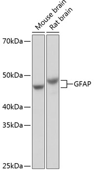
Western blot analysis of various lysates using GFAP Rabbit mAb (A19058) at 1:1000 dilution incubated overnight at 4℃. Secondary antibody: HRP-conjugated Goat anti-Rabbit IgG (H+L) (AS014) at 1:10000 dilution. Lysates/proteins: 25μg per lane. Blocking buffer: 3% nonfat dry milk in TBST. Detection: ECL Basic Kit (RM00020). Exposure time: 1s.

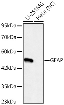
Western blot analysis of various lysates using GFAP Rabbit mAb (A19058)at 1:1000 dilution incubated overnight at 4℃. Secondary antibody: HRP-conjugated Goat anti-Rabbit IgG (H+L) (AS014) at 1:10000 dilution. Lysates/proteins: 25 μg per lane. Blocking buffer: 3% nonfat dry milk in TBST. Detection: ECL Basic Kit (RM00020). Negative control (NC): HeLa Exposure time: 45s.

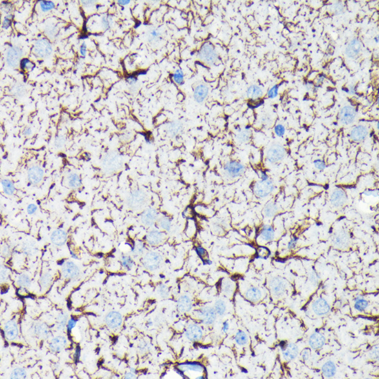
Immunohistochemistry analysis of paraffin-embedded Rat brain using GFAP Rabbit mAb (A19058) at dilution of 1:100 (40x lens). Microwave antigen retrieval performed with 0.01M PBS Buffer (pH 7.2) prior to IHC staining.

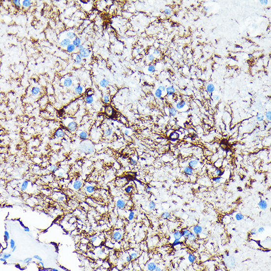
Immunohistochemistry analysis of paraffin-embedded Human brain using GFAP Rabbit mAb (A19058) at dilution of 1:100 (40x lens). Microwave antigen retrieval performed with 0.01M PBS Buffer (pH 7.2) prior to IHC staining.

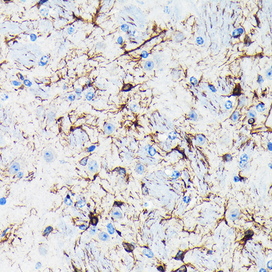
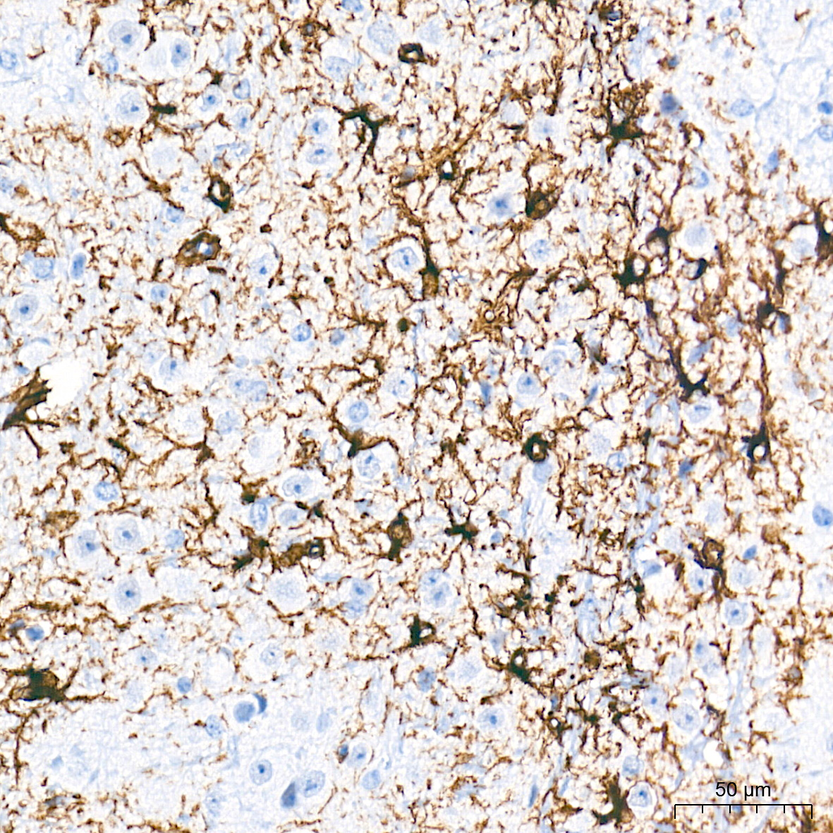
Immunohistochemistry analysis of paraffin-embedded Mouse brain using GFAP Rabbit mAb (A19058) at dilution of 1:100 (40x lens). Microwave antigen retrieval performed with 0.01M PBS Buffer (pH 7.2) prior to IHC staining.

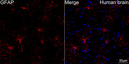
Confocal imaging of paraffin-embedded Human brain tissue using GFAP Rabbit mAb (A19058, dilution 1:1000) followed by a further incubation with Cy3 Goat Anti-Rabbit IgG (H+L) (AS007, dilution 1:500) (Red). DAPI was used for nuclear staining (Blue). High pressure antigen retrieval performed with 0.01M Citrate Buffer (pH 6.0) prior to IF staining. Objective: 40x.

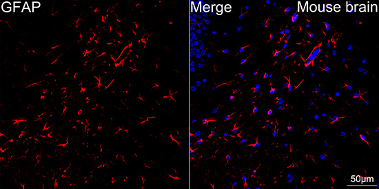
Confocal imaging of paraffin-embedded Mouse brain tissue using GFAP Rabbit mAb (A19058, dilution 1:2000) followed by a further incubation with Cy3 Goat Anti-Rabbit IgG (H+L) (AS007, dilution 1:500) (Red). DAPI was used for nuclear staining (Blue). High pressure antigen retrieval performed with 0.01M Citrate Buffer (pH 6.0) prior to IF staining. Objective: 40x.

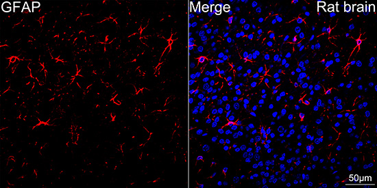
Confocal imaging of paraffin-embedded Rat brain tissue using GFAP Rabbit mAb (A19058, dilution 1:2000) followed by a further incubation with Cy3 Goat Anti-Rabbit IgG (H+L) (AS007, dilution 1:500) (Red). DAPI was used for nuclear staining (Blue). High pressure antigen retrieval performed with 0.01M Citrate Buffer (pH 6.0) prior to IF staining. Objective: 40x.

No documents available.