GFAP Rabbit Monoclonal Antibody

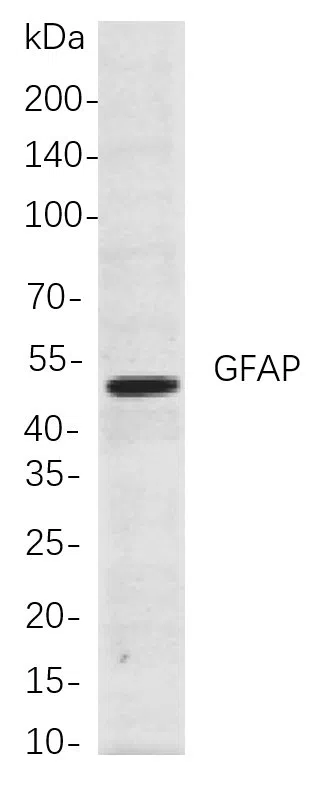
Western blot analysis of lysates from Mouse brain cells, using GFAP Rabbit mAb. The HRP-conjugated Goat anti-Rabbit IgG antibody was used to detect the antibody.

Western blot analysis of lysates from Mouse brain cells, using GFAP Rabbit mAb. The HRP-conjugated Goat anti-Rabbit IgG antibody was used to detect the antibody.

Catalog Number:MABN21476
Conjugate:Unconjugated
Size:200μL
Concentration:1mg/ml
Host:Rabbit
Isotype:IgG,Kappa
Reactivity:Human,Mouse,Rat
Applications:WB 1:1000-1:5000,IHC 1:200-1:1000,ICC/IF 1:200-1:1000,ELISA 1:5000-1:20000,IP 1:50-1:200
Molecular Weight:Calculated MW:50kD;Observed MW:50kD
Purification:Affinity purification
Form:liquid
Synonyms:GFAP;Glial fibrillary acidic protein;GFAP

Cell localization:Cytoplasm.This gene encodes one of the major intermediate filament proteins of mature astrocytes. It is used as a marker to distinguish astrocytes from other glial cells during development. Mutations in this gene cause Alexander disease, a rare disorder of astrocytes in the central nervous system. Alternative splicing results in multiple transcript variants encoding distinct isoforms.

Applications

WB 1:1000-1:5000,IHC 1:200-1:1000,ICC/IF 1:200-1:1000,ELISA 1:5000-1:20000,IP 1:50-1:200

Target Background

Cell localization:Cytoplasm.This gene encodes one of the major intermediate filament proteins of mature astrocytes. It is used as a marker to distinguish astrocytes from other glial cells during development. Mutations in this gene cause Alexander disease, a rare disorder of astrocytes in the central nervous system. Alternative splicing results in multiple transcript variants encoding distinct isoforms.

Synonyms:GFAP;Glial fibrillary acidic protein;GFAP

Storage

Store at 4°C short term. Aliquot and store at -20°C long term. Avoid freeze/thaw cycles.

Buffer

PBS, 50% glycerol, 0.05% Proclin 300, 0.05%protective protein

Western blot analysis of lysates from Mouse brain cells, using GFAP Rabbit mAb. The HRP-conjugated Goat anti-Rabbit IgG antibody was used to detect the antibody.

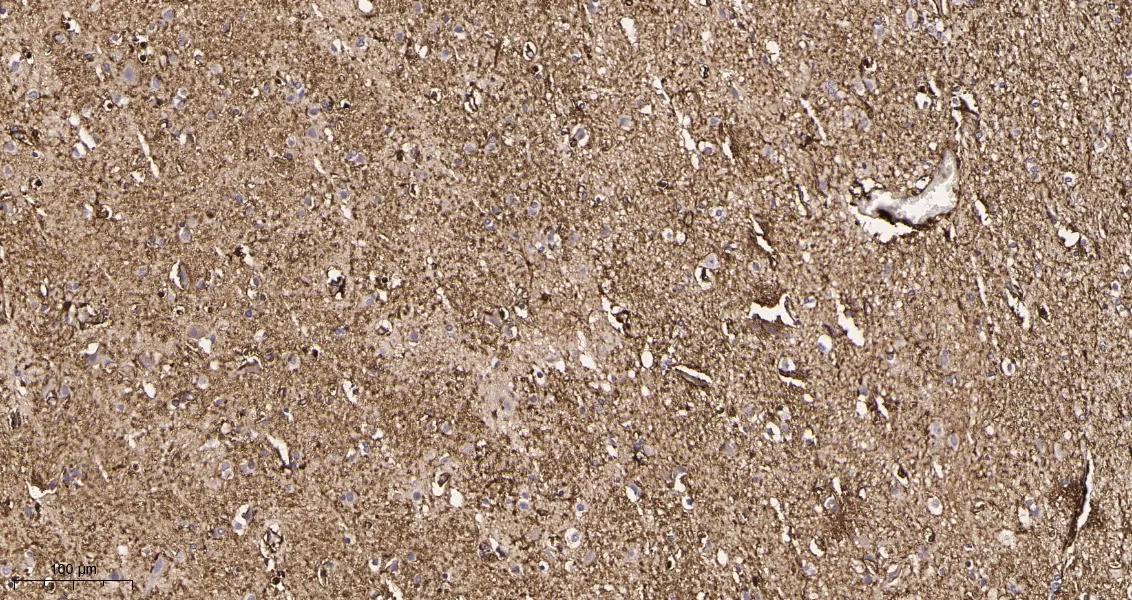
Immunohistochemical analysis of paraffin-embedded Human brain tissue. 1, GFAP Rabbit Monoclonal Antibody was diluted at 1:200(4°C,overnight). 2, EDTA pH 9.0 was used for antibody retrieval(>98°C,20min). 3,Secondary antibody was diluted at 1:200(room temperature, 30min).
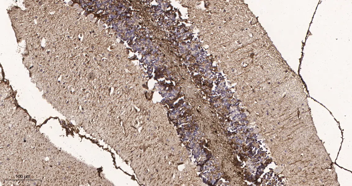
Immunohistochemical analysis of paraffin-embedded Mouse brain tissue. 1, GFAP Rabbit Monoclonal Antibody was diluted at 1:200(4°C,overnight). 2, EDTA pH 9.0 was used for antibody retrieval(>98°C,20min). 3,Secondary antibody was diluted at 1:200(room temperature, 30min).

No documents available.