GLI-3 Rabbit Monoclonal Antibody

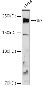
Western blot analysis of extracts of HeLa cells, using Gli3 antibody at 1:1000 dilution.
Secondary antibody: HRP Goat Anti-Rabbit IgG (H+L) at 1:10000 dilution.
Lysates/proteins: 25μg per lane.
Blocking buffer: 3% nonfat dry milk in TBST.
Detection: ECL West Pico  plus.
Exposure time: 180s.

Western blot analysis of extracts of HeLa cells, using Gli3 antibody at 1:1000 dilution. Secondary antibody: HRP Goat Anti-Rabbit IgG (H+L) at 1:10000 dilution. Lysates/proteins: 25μg per lane. Blocking buffer: 3% nonfat dry milk in TBST. Detection: ECL West Pico plus. Exposure time: 180s.

Catalog Number:MAB-94798
Conjugate:Unconjugated
Size:100 ug
Concentration:1mg/ml
Host:Rabbit
Isotype:IgG
Clone:ARC1941
Immunogen:Recombinant fusion protein containing a sequence corresponding to amino acids 1301-1580 of human Gli3
Reactivity:Human, Mouse, Rat
Applications:Western Blot: 1:500 - 1:1000 Immunofluorescence: 1:50 - 1:200 Immunocytcoehmisty: 1:50 - 1:200
Molecular Weight:190 kDa
Purification:Affinity purification
Form:Liquid
Synonyms:PHS; ACLS; GCPS; PAPA; PAPB; PAP-A; PAPA1; PPDIV; GLI3FL; GLI3-190; Gli3

Applications

Western Blot: 1:500 - 1:1000 Immunofluorescence: 1:50 - 1:200 Immunocytcoehmisty: 1:50 - 1:200

Immunogen

Recombinant fusion protein containing a sequence corresponding to amino acids 1301-1580 of human Gli3

Target Background

This gene encodes a protein which belongs to the C2H2-type zinc finger proteins subclass of the Gli family. They are characterized as DNA-binding transcription factors and are mediators of Sonic hedgehog (Shh) signaling. The protein encoded by this gene localizes in the cytoplasm and activates patched Drosophila homolog (PTCH) gene expression. It is also thought to play a role during embryogenesis. Mutations in this gene have been associated with several diseases, including Greig cephalopolysyndactyly syndrome, Pallister-Hall syndrome, preaxial polydactyly type IV, and postaxial polydactyly types A1 and B.

Synonyms:PHS; ACLS; GCPS; PAPA; PAPB; PAP-A; PAPA1; PPDIV; GLI3FL; GLI3-190; Gli3

Storage

Store at -20℃. Avoid freeze / thaw cycles.

Buffer

PBS with 0.02% sodium azide,0.05% BSA,50% glycerol,pH7.3.

Western blot analysis of extracts of HeLa cells, using Gli3 antibody at 1:1000 dilution. Secondary antibody: HRP Goat Anti-Rabbit IgG (H+L) at 1:10000 dilution. Lysates/proteins: 25μg per lane. Blocking buffer: 3% nonfat dry milk in TBST. Detection: ECL West Pico plus. Exposure time: 180s.

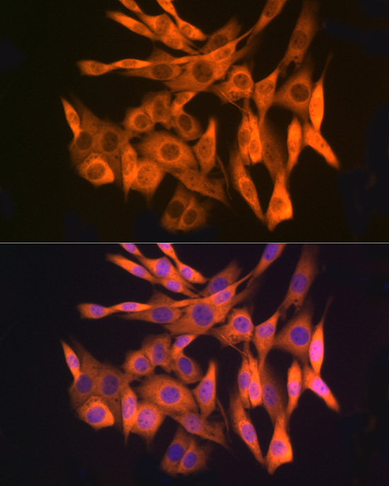
Immunofluorescence analysis of C6 cells using Gli3 Rabbit mAb at dilution of 1:100 (40x lens). Blue: DAPI for nuclear staining.

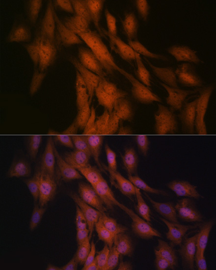
Immunofluorescence analysis of U-2 OS cells using Gli3 Rabbit mAb at dilution of 1:100 (40x lens). Blue: DAPI for nuclear staining.

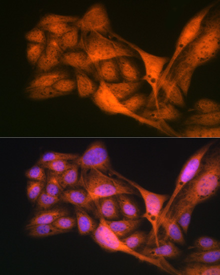
Immunofluorescence analysis of NIH/3T3 cells using Gli3 Rabbit mAb at dilution of 1:100 (40x lens). Blue: DAPI for nuclear staining.

No documents available.