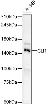
Western blot analysis of lysates from A-549 cells using GLI1 Rabbit  at 1:1000 dilution incubated overnight at 4℃.
Secondary antibody: HRP-conjugated Goat anti-Rabbit IgG (H+L)  at 1:10000 dilution.
Lysates/proteins: 25μg per lane.
Blocking buffer: 3% nonfat dry milk in TBST.
Detection: ECL West Pico Plus
Exposure time: 60s.

Western blot analysis of lysates from A-549 cells using GLI1 Rabbit at 1:1000 dilution incubated overnight at 4℃. Secondary antibody: HRP-conjugated Goat anti-Rabbit IgG (H+L) at 1:10000 dilution. Lysates/proteins: 25μg per lane. Blocking buffer: 3% nonfat dry milk in TBST. Detection: ECL West Pico Plus Exposure time: 60s.

Catalog Number:AB23236
Conjugate:Unconjugated
Size:100 ug
Concentration:1mg/ml
Host:Rb
Isotype:IgG
Clone:POLY
Immunogen:Recombinant fusion protein containing a sequence corresponding to amino acids 1-233 of human GLI1 (NP_005260.1).
Reactivity:Hu, Ms, Rt
Applications:WB, IHC, IF
Molecular Weight:160kDa
Purification:Aff. Pur.
Form:Liquid

This gene encodes a member of the Kruppel family of zinc finger proteins. The encoded transcription factor is activated by the sonic hedgehog signal transduction cascade and regulates stem cell proliferation. The activity and nuclear localization of this protein is negatively regulated by p53 in an inhibitory loop. Multiple transcript variants encoding different isoforms have been found for this gene.

Applications

WB, IHC, IF

Immunogen

Recombinant fusion protein containing a sequence corresponding to amino acids 1-233 of human GLI1 (NP_005260.1).

Target Background

This gene encodes a member of the Kruppel family of zinc finger proteins. The encoded transcription factor is activated by the sonic hedgehog signal transduction cascade and regulates stem cell proliferation. The activity and nuclear localization of this protein is negatively regulated by p53 in an inhibitory loop. Multiple transcript variants encoding different isoforms have been found for this gene.

Storage

Store at -20℃. Avoid freeze / thaw cycles.

Buffer

PBS with 0.05% proclin300,0.05% BSA,50% glycerol,pH7.3.

Western blot analysis of lysates from A-549 cells using GLI1 Rabbit at 1:1000 dilution incubated overnight at 4℃. Secondary antibody: HRP-conjugated Goat anti-Rabbit IgG (H+L) at 1:10000 dilution. Lysates/proteins: 25μg per lane. Blocking buffer: 3% nonfat dry milk in TBST. Detection: ECL West Pico Plus Exposure time: 60s.

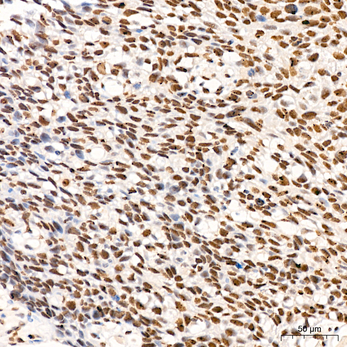
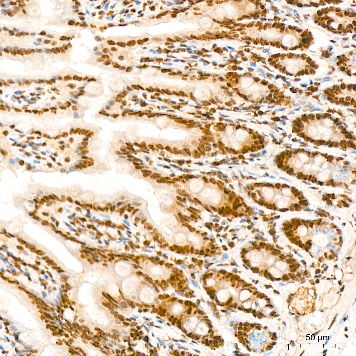
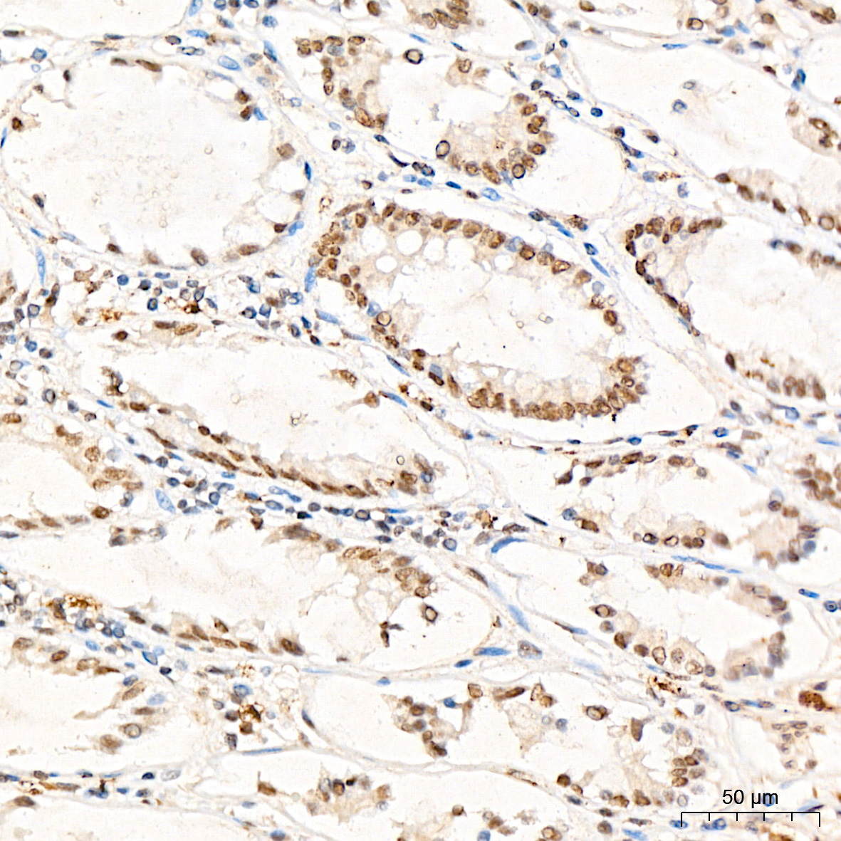
Immunohistochemistry analysis of paraffin-embedded Human colon tissue using GLI1 Rabbit at a dilution of 1:200 (40x lens). High pressure antigen retrieval performed with 0.01M Citrate Buffer (pH 6.0) prior to IHC staining.

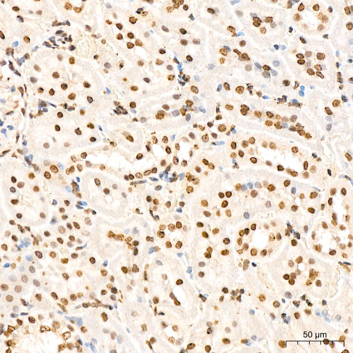
Immunohistochemistry analysis of paraffin-embedded Rat kidney tissue using GLI1 Rabbit mAb at a dilution of 1:200 (40x lens). High pressure antigen retrieval performed with 0.01M Citrate Buffer (pH 6.0) prior to IHC staining.

Immunohistochemistry analysis of paraffin-embedded Human cervix cancer tissue using GLI1 Rabbit at a dilution of 1:200 (40x lens). High pressure antigen retrieval performed with 0.01M Citrate Buffer (pH 6.0) prior to IHC staining.

Immunohistochemistry analysis of paraffin-embedded Mouse colon tissue using GLI1 Rabbit at a dilution of 1:200 (40x lens). High pressure antigen retrieval performed with 0.01M Citrate Buffer (pH 6.0) prior to IHC staining.

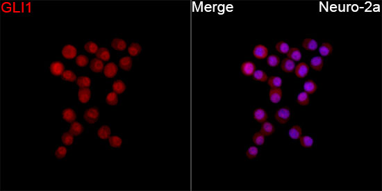
Immunofluorescence analysis of Neuro-2a cells using GLI1 Rabbit mAb at a dilution of 1:100 (40x lens). Secondary antibody: Cy3 Goat Anti-Rabbit IgG (H+L) at 1:500 dilution. Blue: DAPI for nuclear staining.

No documents available.