GM-130 Rabbit Polyclonal Antibody

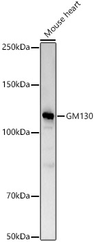
Western blot analysis of Mouse heart, using GM130 antibody at 1:2000 dilution.
Secondary antibody: HRP Goat Anti-Rabbit IgG (H+L) at 1:10000 dilution.
Lysates/proteins: 25μg per lane.
Blocking buffer: 3% nonfat dry milk in TBST.
Detection: ECL West Pico Plus.
Exposure time: 60s.

Western blot analysis of Mouse heart, using GM130 antibody at 1:2000 dilution. Secondary antibody: HRP Goat Anti-Rabbit IgG (H+L) at 1:10000 dilution. Lysates/proteins: 25μg per lane. Blocking buffer: 3% nonfat dry milk in TBST. Detection: ECL West Pico Plus. Exposure time: 60s.

Catalog Number:AB-83480
Conjugate:Unconjugated
Size:100 ug
Concentration:1mg/ml
Host:Rb
Isotype:IgG
Clone:POLY
Immunogen:Recombinant fusion protein containing a sequence corresponding to amino acids 20-300 of human GM130 (
Reactivity:Hu, Ms, Rt
Applications:Western Blot: 1:1000 - 1:5000 Immunohistochemistry (paraffin-embedded tissues): 1:50 - 1:200 Immunofluorescence: 1:100 - 1:500 Immunocytochemistry: 1:100 - 1:500
Molecular Weight:130kDa
Purification:Affinity purification
Form:Liquid
Synonyms:GM130; DEDHMB

Applications

Western Blot: 1:1000 - 1:5000 Immunohistochemistry (paraffin-embedded tissues): 1:50 - 1:200 Immunofluorescence: 1:100 - 1:500 Immunocytochemistry: 1:100 - 1:500

Immunogen

Recombinant fusion protein containing a sequence corresponding to amino acids 20-300 of human GM130 (

Target Background

The Golgi apparatus, which participates in glycosylation and transport of proteins and lipids in the secretory pathway, consists of a series of stacked cisternae (flattened membrane sacs). Interactions between the Golgi and microtubules are thought to be important for the reorganization of the Golgi after it fragments during mitosis. This gene encodes one of the golgins, a family of proteins localized to the Golgi. This encoded protein has been postulated to play roles in the stacking of Golgi cisternae and in vesicular transport. Several alternatively spliced transcript variants of this gene have been described, but the full-length nature of these variants has not been determined.

Synonyms:GM130; DEDHMB

Storage

Store at -20℃. Avoid freeze / thaw cycles.

Buffer

PBS with 0.05% proclin300,50% glycerol,pH7.3.

Western blot analysis of Mouse heart, using GM130 antibody at 1:2000 dilution. Secondary antibody: HRP Goat Anti-Rabbit IgG (H+L) at 1:10000 dilution. Lysates/proteins: 25μg per lane. Blocking buffer: 3% nonfat dry milk in TBST. Detection: ECL West Pico Plus. Exposure time: 60s.

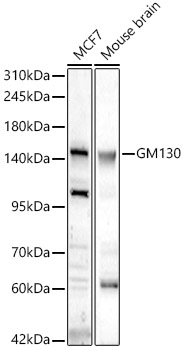
Western blot analysis of various lysates, using GM130 Rabbit pAb at 1:700 dilution. Secondary antibody: HRP Goat Anti-Rabbit IgG (H+L) at 1:10000 dilution. Lysates/proteins: 25ug per lane. Blocking buffer: 3% nonfat dry milk in TBST. Detection: ECL West Pico Plus. Exposure time: 60s.

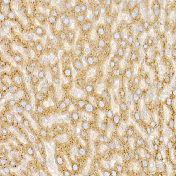
Immunohistochemistry analysis of paraffin-embedded Rat liver using GM130 antibody at dilution of 1:20 (40x lens). Perform high pressure antigen retrieval with 10 mM citrate buffer pH 6.0 before commencing with IHC staining protocol.

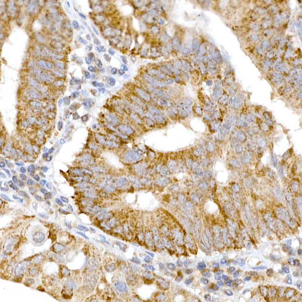
Immunohistochemistry analysis of paraffin-embedded human colon carcinoma using GM130 antibody at dilution of 1:20 (40x lens).Perform high pressure antigen retrieval with 10 mM citrate buffer pH 6.0 before commencing with IHC staining protocol.

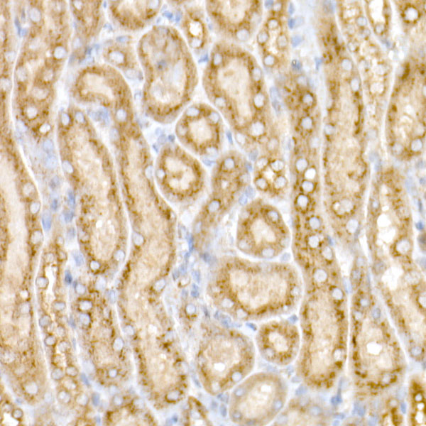
Immunohistochemistry analysis of paraffin-embedded Mouse kidney using GM130 antibody at dilution of 1:20 (40x lens).Perform high pressure antigen retrieval with 10 mM citrate buffer pH 6.0 before commencing with IHC staining protocol.

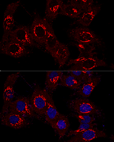
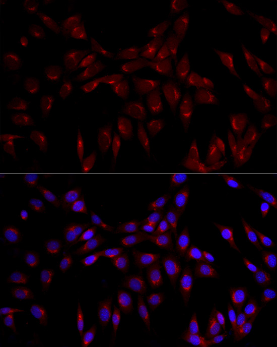
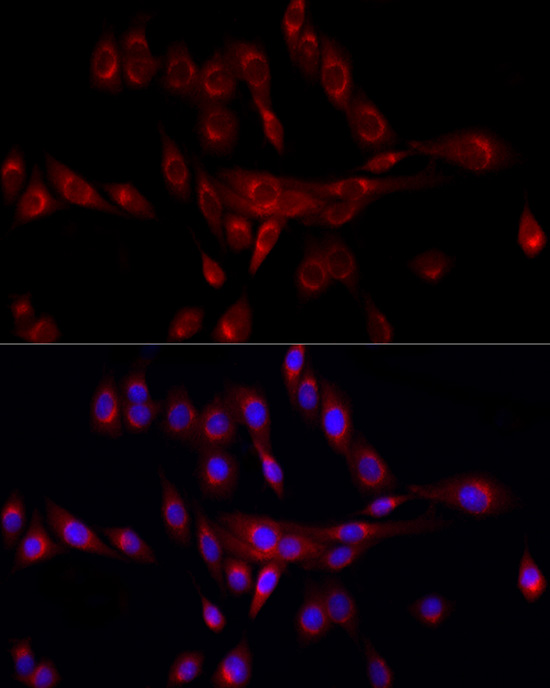
Confocal immunofluorescence analysis of Hela cells using GM130 antibody at dilution of 1:400. Blue: DAPI for nuclear staining.

Immunofluorescence analysis of NIH/3T3 cells using GM130 Rabbit pAb at dilution of 1:100 (40x lens). Blue: DAPI for nuclear staining.

Immunofluorescence analysis of PC-12 cells using GM130 Rabbit pAb at dilution of 1:100 (40x lens). Blue: DAPI for nuclear staining.

No documents available.