HA Tag Mouse Monoclonal Antibody

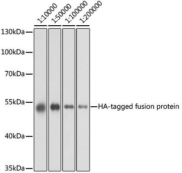
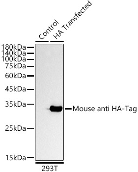
Western blot analysis of over-expressed HA-tagged protein in 293T cell using HA-tag antibody at different dilution. Each lane was loaded with 2 ug cell lysate.
Secondary antibody: HRP Goat Anti-Mouse IgG (H+L) t 1:10000 dilution.
Blocking buffer: 3% nonfat dry milk in TBST.
Detection: ECL West Pico Plus.
Exposure time: 1s.

Western blot analysis of over-expressed HA-tagged protein in 293T cell using HA-tag antibody at different dilution. Each lane was loaded with 2 ug cell lysate. Secondary antibody: HRP Goat Anti-Mouse IgG (H+L) t 1:10000 dilution. Blocking buffer: 3% nonfat dry milk in TBST. Detection: ECL West Pico Plus. Exposure time: 1s.

Catalog Number:MAB-91878
Conjugate:Unconjugated
Size:100 ug
Concentration:1mg/mL
Host:Ms
Isotype:IgG1,Kappa
Clone:2D10
Immunogen:A synthetic peptide corresponding to HA tag.
Reactivity:All Species
Applications:Western Blot: 1:2000 - 1:10000 Immunofluorescence:1:50 - 1:200 Immunocytochemistry: 1:50 - 1:200 Immunoprecipitation: 0.5μg-4μg antibody for 200μg-400μg extracts of whole cells
Purification:Affinity purification
Form:Liquid
Synonyms:HA;HA tag;HA-tag

Applications

Western Blot: 1:2000 - 1:10000 Immunofluorescence:1:50 - 1:200 Immunocytochemistry: 1:50 - 1:200 Immunoprecipitation: 0.5μg-4μg antibody for 200μg-400μg extracts of whole cells

Immunogen

A synthetic peptide corresponding to HA tag.
Target Background

Protein tags are peptide sequences genetically grafted onto a recombinant protein. Often these tags are removable by chemical agents or by enzymatic means, such as proteolysis or intein splicing. Tags are attached to proteins for various purposes.Epitope tags are short peptide sequences which are chosen because high-affinity antibodies can be reliably produced in many different species. These are usually derived from viral genes, which explain their high immunoreactivity. Epitope tags include V5-tag, Myc-tag, HA-tag and NE-tag. These tags are particularly useful for western blotting, immunofluorescence and immunoprecipitation experiments, although they also find use in antibody purification.

Synonyms:HA;HA tag;HA-tag

Storage

Store at -20°C for 1 year. Avoid repeated freeze and thaw cycles.

Buffer

PBS with 0.01% thimerosal,50% glycerol,pH7.3.

Western blot analysis of over-expressed HA-tagged protein in 293T cell using HA-tag antibody at different dilution. Each lane was loaded with 2 ug cell lysate. Secondary antibody: HRP Goat Anti-Mouse IgG (H+L) t 1:10000 dilution. Blocking buffer: 3% nonfat dry milk in TBST. Detection: ECL West Pico Plus. Exposure time: 1s.

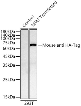
Western blot analysis of lysates from 293T cells, using Mouse anti HA-Tag mAb at 1:5000 dilution. Secondary antibody: HRP Goat Anti-Mouse IgG (H+L) at 1:10000 dilution. Lysates/proteins: 25μg per lane. Blocking buffer: 3% nonfat dry milk in TBST. Detection: ECL West Pico Plus. Exposure time: 10s.

Western blot analysis of lysates from 293T cells, using Mouse anti HA-Tag mAb at 1:10000 dilution. Secondary antibody: HRP Goat Anti-Mouse IgG (H+L)at 1:10000 dilution. Lysates/proteins: 25μg per lane. Blocking buffer: 3% nonfat dry milk in TBST. Detection: ECL West Pico Plus. Exposure time: 180s.

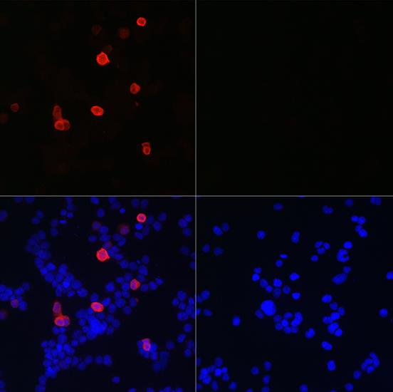
Immunofluorescence analysis of 293T cells transfected with HA-Tag fusion protein and untreated 293T cells use Mouse anti HATag mAb at dilution of 1:50 (40xlens). Blue: DAPI for nuclear staining.

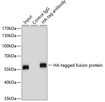
Immunoprecipitation of over-expressed HA-tagged protein in 293T cells using HAtag antibody . A mock served as negative control and over-expressed 293T cell lysate served as positive control.

No documents available.