Hepatocyte Growth Factor

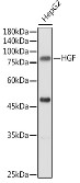
Western blot analysis of extracts of HepG2 cells, using HGF antibody at 1:500 dilution.   Secondary antibody: HRP Goat Anti-Rabbit IgG (H+L) at 1:10000 dilution.  
Lysates/proteins: 25ug per lane.     Blocking buffer: 3% nonfat dry milk in TBST.  Detection: ECL West Pico Plus.  
Exposure time: 30s.

Western blot analysis of extracts of HepG2 cells, using HGF antibody at 1:500 dilution. Secondary antibody: HRP Goat Anti-Rabbit IgG (H+L) at 1:10000 dilution. Lysates/proteins: 25ug per lane. Blocking buffer: 3% nonfat dry milk in TBST. Detection: ECL West Pico Plus. Exposure time: 30s.

Catalog Number:AB-90164
Size:100 ug
Concentration:1mg/ml
Host:Rb
Isotype:IgG
Clone:POLY
Immunogen:Recombinant fusion protein containing a sequence corresponding to amino acids 30-285 of human HGF.
Reactivity:Hu, Ms, Rt
Applications:Western Blot: 1:500-1:2000 Immunohistochemistry: 1:50 - 1:100
Molecular Weight:83kDa
Purification:Aff. Pur.
Form:Liquid
Synonyms:HGF;DFNB39;F-TCF;HGFB;HPTA;SF

Applications

Western Blot: 1:500-1:2000 Immunohistochemistry: 1:50 - 1:100

Immunogen

Recombinant fusion protein containing a sequence corresponding to amino acids 30-285 of human HGF.
Target Background

This gene encodes a protein that binds to the hepatocyte growth factor receptor to regulate cell growth, cell motility and morphogenesis in numerous cell and tissue types. Alternative splicing results in multiple transcript variants, at least one of which encodes a preproprotein that is proteolytically processed to generate alpha and beta chains, which form the mature heterodimer. This protein is secreted by mesenchymal cells and acts as a multi-functional cytokine on cells of mainly epithelial origin. This protein also plays a role in angiogenesis, tumorogenesis, and tissue regeneration. Although the encoded protein is a member of the peptidase S1 family of serine proteases, it lacks peptidase activity. Mutations in this gene are associated with non syndromic hearing loss.

Synonyms:HGF;DFNB39;F-TCF;HGFB;HPTA;SF

Storage

Store at -20℃. Avoid freeze / thaw cycles.

Buffer

PBS with 0.02% sodium azide,50% glycerol,pH7.3.

Western blot analysis of extracts of HepG2 cells, using HGF antibody at 1:500 dilution. Secondary antibody: HRP Goat Anti-Rabbit IgG (H+L) at 1:10000 dilution. Lysates/proteins: 25ug per lane. Blocking buffer: 3% nonfat dry milk in TBST. Detection: ECL West Pico Plus. Exposure time: 30s.

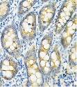
Immunohistochemistry of paraffin embedded rat intestine using HGF antibody at dilution of 1:100 (40x lens).

No documents available.